3OB1 image
Deposition Date 2010-08-06
Release Date 2010-08-18
Last Version Date 2024-11-20
Entry Detail
PDB ID:
3OB1
Title:
Crystal structure of c-Cbl TKB domain in complex with double phosphorylated Spry2 peptide
Biological Source:
Source Organism:
Homo sapiens (Taxon ID: 9606)
Host Organism:
Method Details:
Experimental Method:
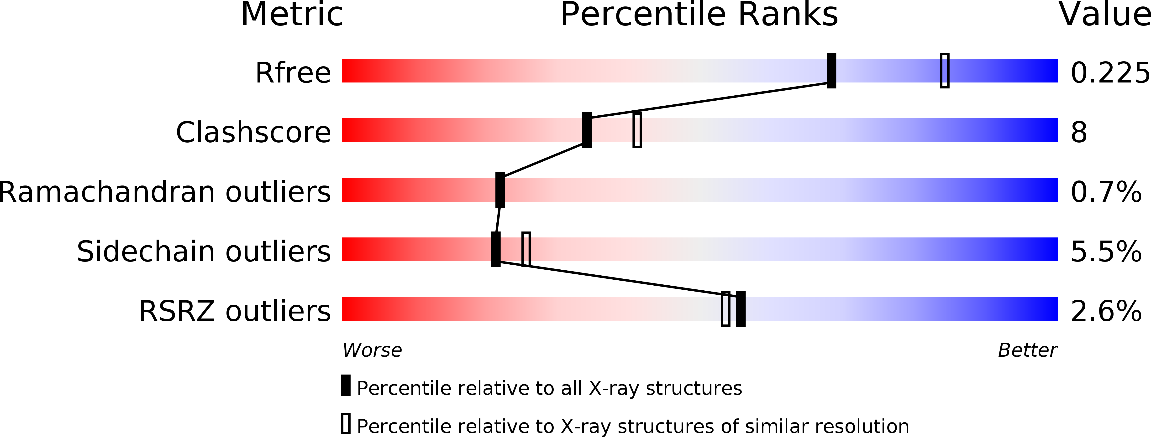
Resolution:
2.20 Å
R-Value Free:
0.22
R-Value Work:
0.18
R-Value Observed:
0.18
Space Group:
P 6
Macromolecular Entities
Polymer Type:polypeptide(L)
Molecule:12-meric peptide from Protein sprouty homolog 2
Gene (Uniprot):SPRY2
Chain IDs:A
Chain Length:12
Number of Molecules:1
Biological Source:Homo sapiens
Polymer Type:polypeptide(L)
Molecule:E3 ubiquitin-protein ligase CBL
Gene (Uniprot):CBL
Chain IDs:B
Chain Length:329
Number of Molecules:1
Biological Source:Homo sapiens
Modified Residue
Compound ID Chain ID Parent Comp ID Details 2D Image
PTR A TYR O-PHOSPHOTYROSINE
TPO A THR PHOSPHOTHREONINE
Primary Citation
Additional serine/threonine phosphorylation reduces binding affinity but preserves interface topography of substrate proteins to the c-Cbl TKB domain
Plos One 5 e12819 e12819 (2010)
PMID: 20877636 DOI: 10.1371/journal.pone.0012819

Abstact

The E3-ubiquitin ligase, c-Cbl, is a multi-functional scaffolding protein that plays a pivotal role in controlling cell phenotype. As part of the ubiquitination and downregulation process, c-Cbl recognizes targets, such as tyrosine kinases and the Sprouty proteins, by binding to a conserved (NX/R)pY(S/T)XXP motif via its uniquely embedded SH2 domain (TKB domain). We previously outlined the mode of binding between the TKB domain and various substrate peptide motifs, including epidermal growth factor receptor (EGFR) and Sprouty2 (Spry2), and demonstrated that an intrapetidyl hydrogen bond forms between the (pY-1) arginine or (pY-2) asparagine and the phosphorylated tyrosine, which is crucial for binding. Recent reports demonstrated that, under certain types of stimulation, the serine/threonine residues at the pY+1 and/or pY+2 positions within this recognition motif of EGFR and Sprouty2 may be endogenously phosphorylated. Using structural and binding studies, we sought to determine whether this additional phosphorylation could affect the binding of the TKB domain to these peptides and consequently, whether the type of stimulation can dictate the degree to which substrates bind to c-Cbl. Here, we show that additional phosphorylation significantly reduces the binding affinity between the TKB domain and its target proteins, EGFR and Sprouty2, as compared to peptides bearing a single tyrosine phosphorylation. The crystal structure indicates that this is accomplished with minimal changes to the essential intrapeptidyl bond and that the reduced strength of the interaction is due to the charge repulsion between c-Cbl and the additional phosphate group. This obvious reduction in binding affinity, however, indicates that Cbl's interactions with its TKB-centered binding partners may be more favorable in the absence of Ser/Thr phosphorylation, which is stimulation and context specific in vivo. These results demonstrate the importance of understanding the environment in which certain residues are phosphorylated, and the necessity of including this in structural investigations.

Legend

Protein

Chemical

Disease

Primary Citation of related structures