Deposition Date
2010-08-05
Release Date
2011-05-25
Last Version Date
2023-09-06
Entry Detail
PDB ID:
3OAA
Keywords:
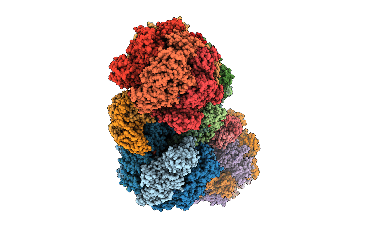
Title:
Structure of the E.coli F1-ATP synthase inhibited by subunit Epsilon
Biological Source:
Source Organism(s):
Escherichia coli DH1 (Taxon ID: 536056)
Expression System(s):
Method Details:
Experimental Method:
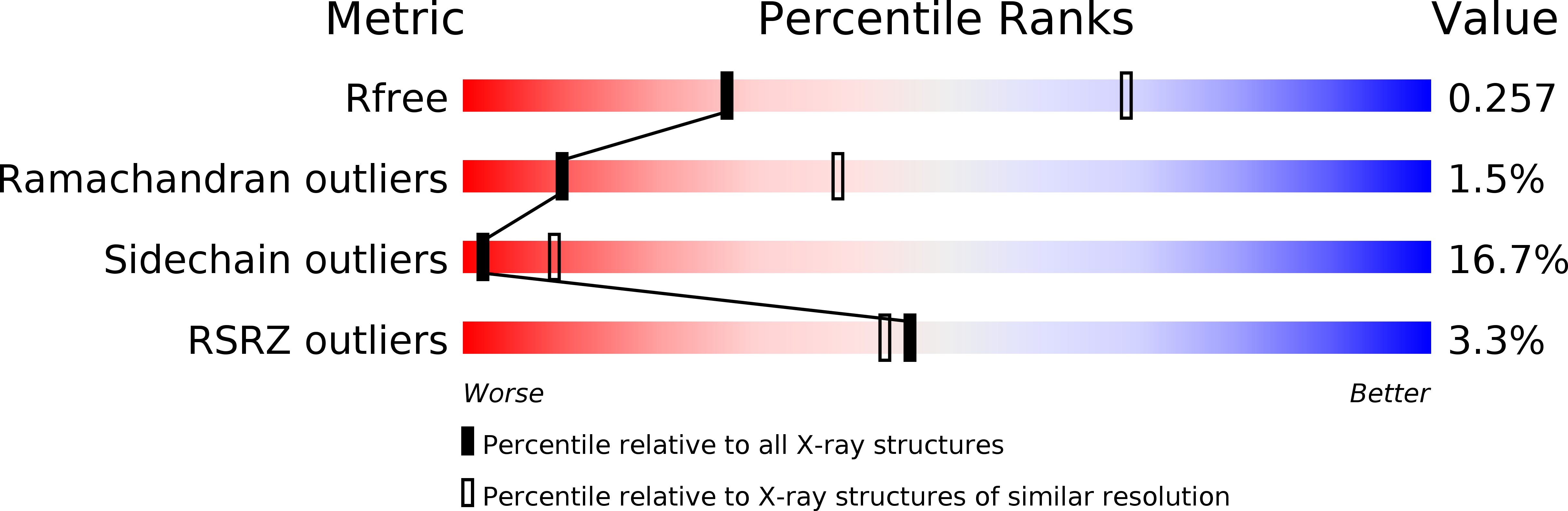
Resolution:
3.26 Å
R-Value Free:
0.26
R-Value Work:
0.24
R-Value Observed:
0.24
Space Group:
C 1 2 1