3O9X image
Deposition Date 2010-08-04
Release Date 2010-11-10
Last Version Date 2023-09-06
Entry Detail
PDB ID:
3O9X
Title:
Structure of the E. coli antitoxin MqsA (YgiT/b3021) in complex with its gene promoter
Biological Source:
Source Organism:
Host Organism:
Method Details:
Experimental Method:
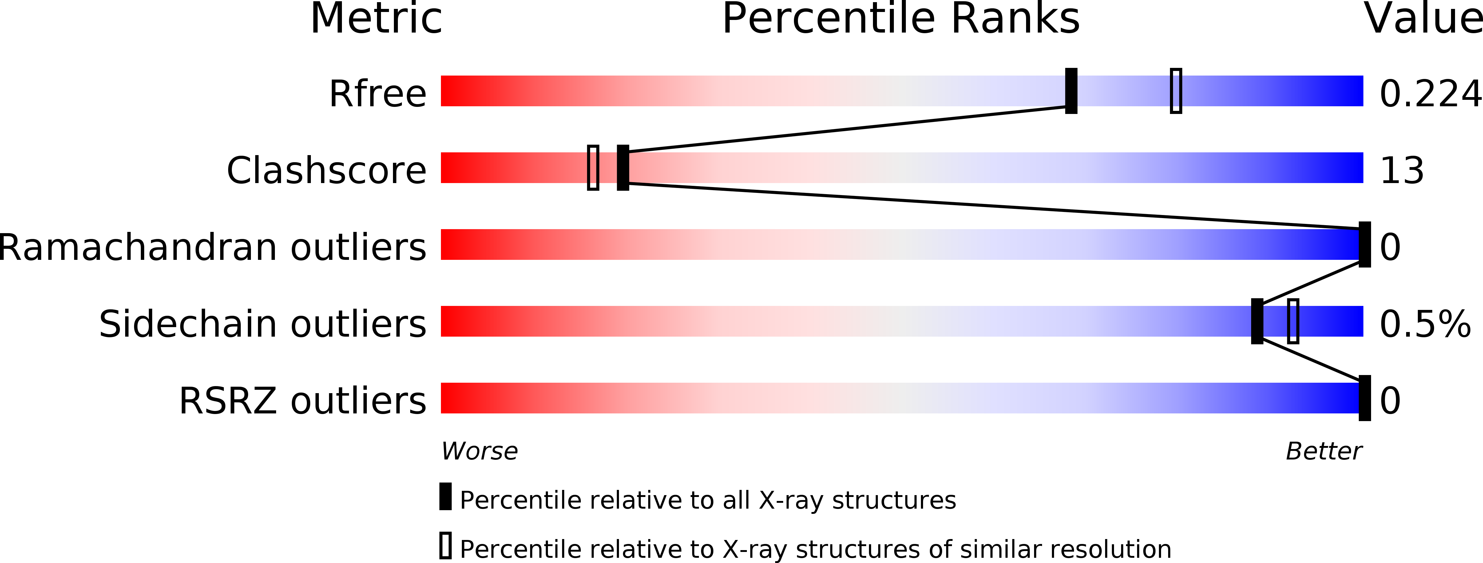
Resolution:
2.10 Å
R-Value Free:
0.22
R-Value Work:
0.17
R-Value Observed:
0.18
Space Group:
P 41
Macromolecular Entities
Structures with similar UniProt ID
Protein Blast
Polymer Type:polypeptide(L)
Molecule:Uncharacterized HTH-type transcriptional regulator ygiT
Gene (Uniprot):mqsA
Chain IDs:A, B
Chain Length:133
Number of Molecules:2
Biological Source:Escherichia coli
Polymer Type:polydeoxyribonucleotide
Molecule:DNA (26-MER)
Chain IDs:D (auth: E)
Chain Length:26
Number of Molecules:1
Biological Source:
Polymer Type:polydeoxyribonucleotide
Molecule:DNA (26-MER)
Chain IDs:C (auth: F)
Chain Length:26
Number of Molecules:1
Biological Source:
Primary Citation
Structure of the Escherichia coli Antitoxin MqsA (YgiT/b3021) Bound to Its Gene Promoter Reveals Extensive Domain Rearrangements and the Specificity of Transcriptional Regulation.
J.Biol.Chem. 286 2285 2296 (2011)
PMID: 21068382 DOI: 10.1074/jbc.M110.172643

Abstact

Bacterial cultures, especially biofilms, produce a small number of persister cells, a genetically identical subpopulation of wild type cells that are metabolically dormant, exhibit multidrug tolerance, and are highly enriched in bacterial toxins. The gene most highly up-regulated in Escherichia coli persisters is mqsR, a ribonuclease toxin that, along with mqsA, forms a novel toxin·antitoxin (TA) system. Like all known TA systems, both the MqsR·MqsA complex and MqsA alone regulate their own transcription. Despite the importance of TA systems in persistence and biofilms, very little is known about how TA modules, and antitoxins in particular, bind and recognize DNA at a molecular level. Here, we report the crystal structure of MqsA bound to a 26-bp fragment from the mqsRA promoter. We show that MqsA binds DNA predominantly via its C-terminal helix-turn-helix domain, with direct binding of recognition helix residues Asn(97) and Arg(101) to the DNA major groove. Unexpectedly, the structure also revealed that the MqsA N-terminal domain interacts with the DNA phosphate backbone. This results in a more than 105° rotation of the N-terminal domains between the free and complexed states, an unprecedented rearrangement for an antitoxin. The structure also shows that MqsA bends the DNA by more than 55° in order to achieve symmetrical binding. Finally, using a combination of biochemical and NMR studies, we show that the DNA sequence specificity of MqsA is mediated by direct readout.

Legend

Protein

Chemical

Disease

Primary Citation of related structures
Feedback Form
Name
Email
Institute
Feedback