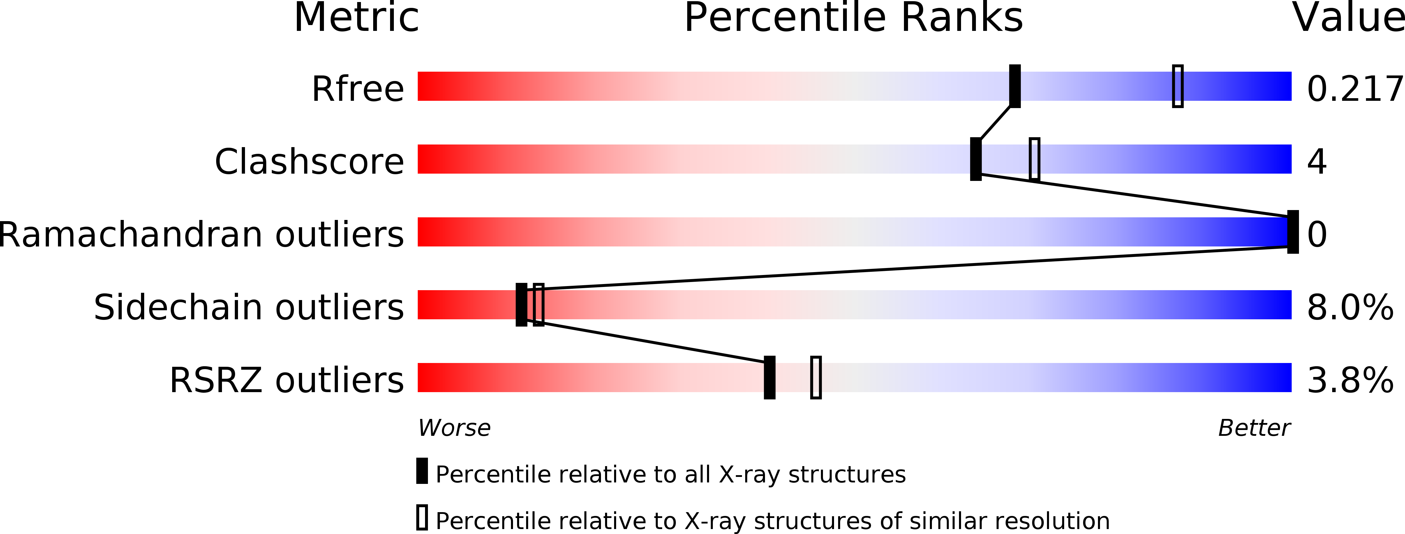
3O8W image
Deposition Date 2010-08-03
Release Date 2011-02-09
Last Version Date 2023-09-06
Entry Detail
PDB ID:
3O8W
Title:
Archaeoglobus fulgidus GlnK1
Biological Source:
Source Organism:
Host Organism:
Method Details:
Experimental Method:
Resolution:
2.28 Å
R-Value Free:
0.22
R-Value Work:
0.17
R-Value Observed:
0.17
Space Group:
I 21 3
Macromolecular Entities
Structures with similar UniProt ID
Protein Blast
Polymer Type:polypeptide(L)
Molecule:Nitrogen regulatory protein P-II (GlnB-1)
Gene (Uniprot):glnK1
Chain IDs:A
Chain Length:118
Number of Molecules:1
Biological Source:Archaeoglobus fulgidus
Ligand Molecules
Primary Citation
Structure of GlnK1, a signalling protein from Archaeoglobus fulgidus.
Acta Crystallogr.,Sect.F 67 178 181 (2011)
PMID: 21301082 DOI: 10.1107/S1744309110047482

Abstact

GlnB and GlnK are ancient signalling proteins that play a crucial role in the regulation of nitrogen assimilation. Both protein types can be present in the same genome as either single or multiple copies. However, the gene product of glnK is always found in an operon together with an amt gene encoding an ammonium-transport (Amt) protein. Complex formation between GlnK and Amt blocks ammonium uptake and depends on the nitrogen level in the cell, which is regulated through the binding of specific effector molecules to GlnK. In particular, an ammonium shock to a cell culture previously starved in this nitrogen source or the binding of ATP to purified GlnK can stimulate effective complex formation. While the binding of ATP/ADP and 2-oxoglutarate (as a signal for low intracellular nitrogen) to GlnK have been reported and several GlnB/K protein structures are available, essential functional questions remain unanswered. Here, the crystal structure of A. fulgidus GlnK1 at 2.28 Å resolution and a comparison with the crystal structures of other GlnK proteins, in particular with that of its paralogue GlnK2 from the same organism, is reported.

Legend

Protein

Chemical

Disease

Primary Citation of related structures