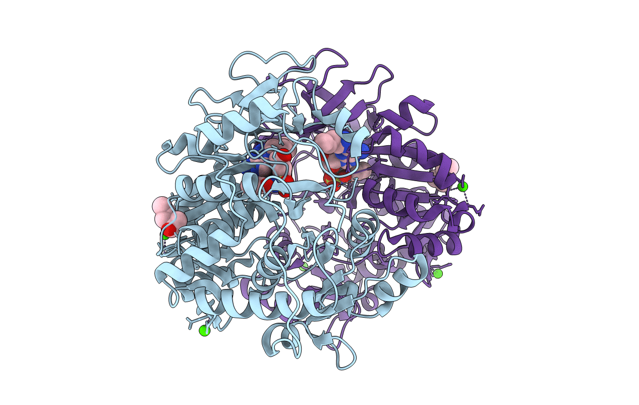
3O83 image
Entry Detail
PDB ID:
3O83
Keywords:
Title:
Structure of BasE N-terminal domain from Acinetobacter baumannii bound to 2-(4-n-dodecyl-1,2,3-triazol-1-yl)-5'-O-[N-(2-hydroxybenzoyl)sulfamoyl]adenosine
Biological Source:
Source Organism:
Host Organism:
PDB Version:
Deposition Date:
2010-08-02
Release Date:
2010-10-06
Method Details:
Experimental Method:
Resolution:
1.90 Å
R-Value Free:
0.21
R-Value Work:
0.19
R-Value Observed:
0.19
Space Group:
P 21 21 21
Macromolecular Entities
Polymer Type:polypeptide(L)
Description:Peptide arylation enzyme
Mutations:P45L
Chain IDs:A, B
Chain Length:544
Number of Molecules:2
Biological Source:Acinetobacter baumannii
Primary Citation
Biochemical and structural characterization of bisubstrate inhibitors of BasE, the self-standing nonribosomal peptide synthetase adenylate-forming enzyme of acinetobactin synthesis.
Biochemistry 49 9292 9305 (2010)
PMID: 20853905 DOI: 10.1021/bi101226n

Abstact

The human pathogen Acinetobacter baumannii produces a siderophore called acinetobactin that is derived from one molecule each of threonine, histidine, and 2,3-dihydroxybenzoic acid (DHB). The activity of several nonribosomal peptide synthetase (NRPS) enzymes is used to combine the building blocks into the final molecule. The acinetobactin synthesis pathway initiates with a self-standing adenylation enzyme, BasE, that activates the DHB molecule and covalently transfers it to the pantetheine cofactor of an aryl-carrier protein of BasF, a strategy that is shared with many siderophore-producing NRPS clusters. In this reaction, DHB reacts with ATP to form the aryl adenylate and pyrophosphate. In a second partial reaction, the DHB is transferred to the carrier protein. Inhibitors of BasE and related enzymes have been identified that prevent growth of bacteria on iron-limiting media. Recently, a new inhibitor of BasE has been identified via high-throughput screening using a fluorescence polarization displacement assay. We present here biochemical and structural studies to examine the binding mode of this inhibitor. The kinetics of the wild-type BasE enzyme is shown, and inhibition studies demonstrate that the new compound exhibits competitive inhibition against both ATP and 2,3-dihydroxybenzoate. Structural examination of BasE bound to this inhibitor illustrates a novel binding mode in which the phenyl moiety partially fills the enzyme pantetheine binding tunnel. Structures of rationally designed bisubstrate inhibitors are also presented.

Legend

Protein

Chemical

Disease

Primary Citation of related structures