3O7U image
Deposition Date 2010-07-31
Release Date 2011-06-15
Last Version Date 2023-10-04
Entry Detail
PDB ID:
3O7U
Title:
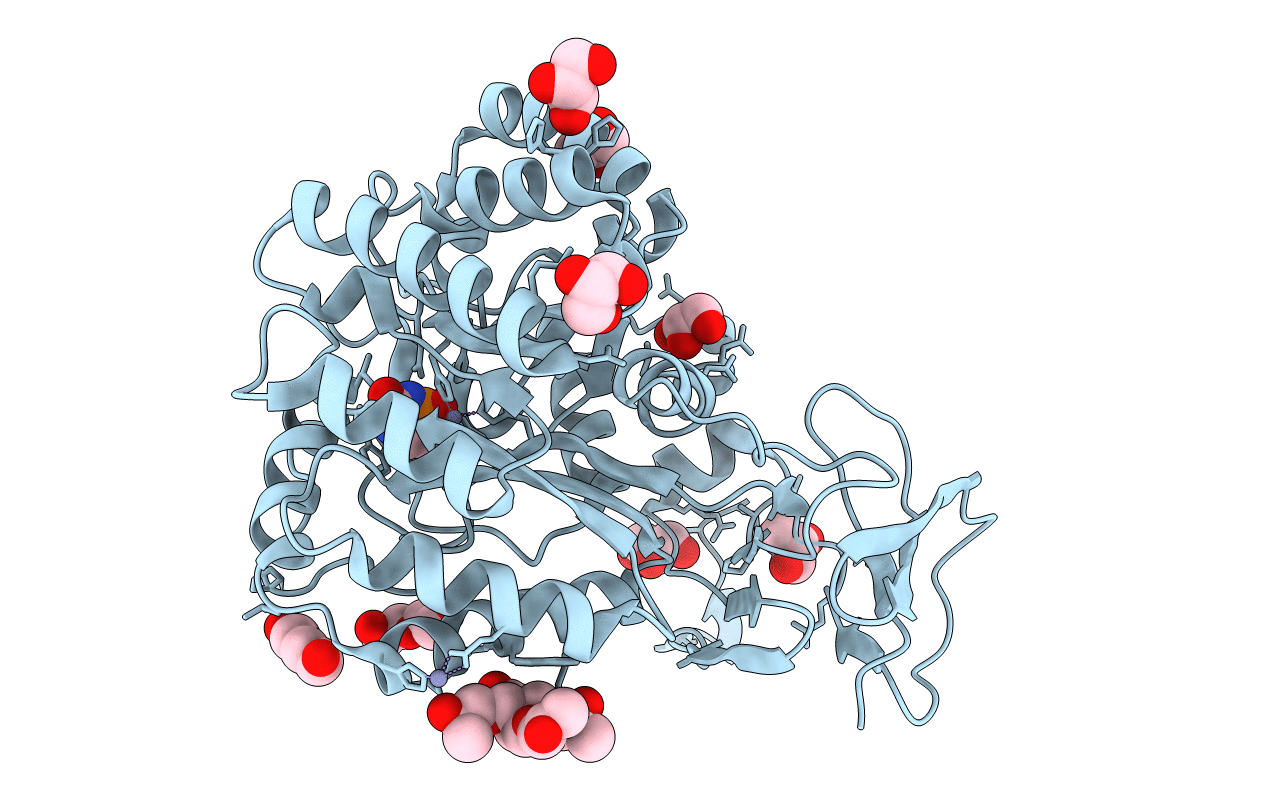
Crystal structure of Cytosine Deaminase from Escherichia Coli complexed with zinc and phosphono-cytosine
Biological Source:
Source Organism:
Escherichia coli (Taxon ID: 562)
Host Organism:
Method Details:
Experimental Method:
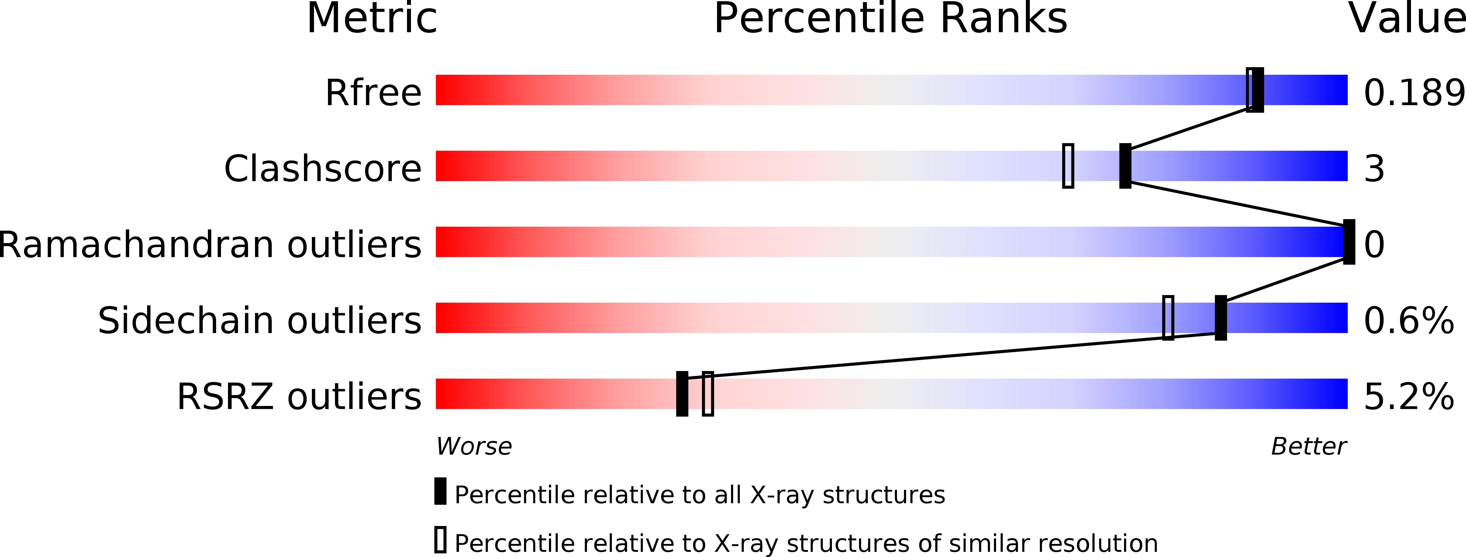
Resolution:
1.71 Å
R-Value Free:
0.19
R-Value Work:
0.17
R-Value Observed:
0.17
Space Group:
H 3 2
Macromolecular Entities
Structures with similar UniProt ID
Protein Blast
Polymer Type:polypeptide(L)
Molecule:Cytosine deaminase
Chain IDs:A
Chain Length:427
Number of Molecules:1
Biological Source:Escherichia coli
Primary Citation
Three-dimensional structure and catalytic mechanism of Cytosine deaminase.
Biochemistry 50 5077 5085 (2011)
PMID: 21545144 DOI: 10.1021/bi200483k

Abstact

Cytosine deaminase (CDA) from E. coli is a member of the amidohydrolase superfamily. The structure of the zinc-activated enzyme was determined in the presence of phosphonocytosine, a mimic of the tetrahedral reaction intermediate. This compound inhibits the deamination of cytosine with a K(i) of 52 nM. The zinc- and iron-containing enzymes were characterized to determine the effect of the divalent cations on activation of the hydrolytic water. Fe-CDA loses activity at low pH with a kinetic pK(a) of 6.0, and Zn-CDA has a kinetic pK(a) of 7.3. Mutation of Gln-156 decreased the catalytic activity by more than 5 orders of magnitude, supporting its role in substrate binding. Mutation of Glu-217, Asp-313, and His-246 significantly decreased catalytic activity supporting the role of these three residues in activation of the hydrolytic water molecule and facilitation of proton transfer reactions. A library of potential substrates was used to probe the structural determinants responsible for catalytic activity. CDA was able to catalyze the deamination of isocytosine and the hydrolysis of 3-oxauracil. Large inverse solvent isotope effects were obtained on k(cat) and k(cat)/K(m), consistent with the formation of a low-barrier hydrogen bond during the conversion of cytosine to uracil. A chemical mechanism for substrate deamination by CDA was proposed.

Legend

Protein

Chemical

Disease

Primary Citation of related structures