3O6R image
Entry Detail
PDB ID:
3O6R
Keywords:
Title:
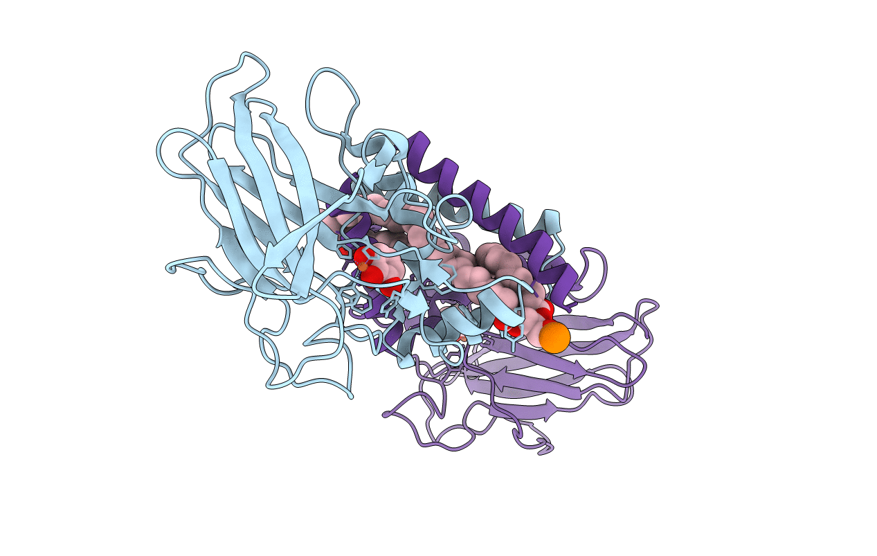
Crystal Structure of 4-Chlorocatechol Dioxygenase from Rhodococcus opacus 1CP in complex with pyrogallol
Biological Source:
Source Organism:
PDB Version:
Deposition Date:
2010-07-29
Release Date:
2011-08-17
Method Details:
Experimental Method:
Resolution:
2.60 Å
R-Value Free:
0.31
R-Value Work:
0.22
R-Value Observed:
0.22
Space Group:
P 63 2 2
Macromolecular Entities
Polymer Type:polypeptide(L)
Description:Chlorocatechol 1,2-dioxygenase
Chain IDs:A, B
Chain Length:257
Number of Molecules:2
Biological Source:RHODOCOCCUS OPACUS
Primary Citation
X-ray structures of 4-chlorocatechol 1,2-dioxygenase adducts with substituted catechols: new perspectives in the molecular basis of intradiol ring cleaving dioxygenases specificity.
J. Struct. Biol. 181 274 282 (2013)
PMID: 23261399 DOI: 10.1016/j.jsb.2012.11.007

Abstact

The crystallographic structures of 4-chlorocatechol 1,2-dioxygenase (4-CCD) complexes with 3,5-dichlorocatechol, protocatechuate (3,4-dihydroxybenzoate), hydroxyquinol (benzen-1,2,4-triol) and pyrogallol (benzen-1,2,3-triol), which act as substrates or inhibitors of the enzyme, have been determined and analyzed. 4-CCD from the Gram-positive bacterium Rhodococcus opacus 1CP is a Fe(III) ion containing enzyme specialized in the aerobic biodegradation of chlorocatechols. The structures of the 4-CCD complexes show that the catechols bind the catalytic iron ion in a bidentate mode displacing Tyr169 and the benzoate ion (found in the native enzyme structure) from the metal coordination sphere, as found in other adducts of intradiol dioxygenases with substrates. The analysis of the present structures allowed to identify the residues selectively involved in recognition of the diverse substrates. Furthermore the structural comparison with the corresponding complexes of catechol 1,2-dioxygenase from the same Rhodococcus strain (Rho-1,2-CTD) highlights significant differences in the binding of the tested catechols to the active site of the enzyme, particularly in the orientation of the aromatic ring substituents. As an example the 3-substituted catechols are bound with the substituent oriented towards the external part of the 4-CCD active site cavity, whereas in the Rho-1,2-CTD complexes the 3-substituents were placed in the internal position. The present crystallographic study shed light on the mechanism that allows substrate recognition inside this class of high specific enzymes involved in the biodegradation of recalcitrant pollutants.

Legend

Protein

Chemical

Disease

Primary Citation of related structures