Deposition Date
2010-07-28
Release Date
2010-11-24
Last Version Date
2024-11-06
Entry Detail
PDB ID:
3O65
Keywords:
Title:
Crystal structure of a Josephin-ubiquitin complex: Evolutionary restraints on ataxin-3 deubiquitinating activity
Biological Source:
Source Organism(s):
Homo sapiens (Taxon ID: 9606)
Expression System(s):
Method Details:
Experimental Method:
Resolution:
2.70 Å
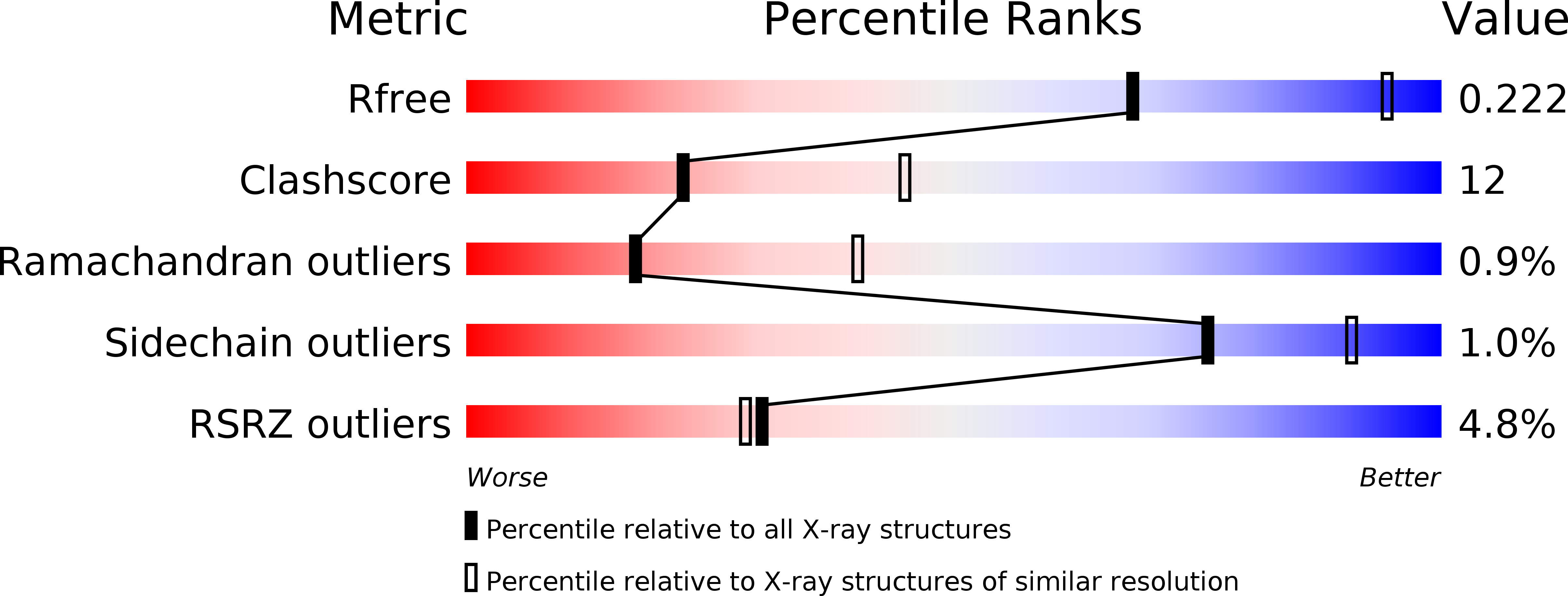
R-Value Free:
0.22
R-Value Work:
0.17
R-Value Observed:
0.17
Space Group:
P 3 2 1