Deposition Date
2010-07-28
Release Date
2011-07-13
Last Version Date
2024-02-21
Entry Detail
PDB ID:
3O5S
Keywords:
Title:
Crystal Structure of the endo-beta-1,3-1,4 glucanase from Bacillus subtilis (strain 168)
Biological Source:
Source Organism(s):
Bacillus subtilis (Taxon ID: 1423)
Expression System(s):
Method Details:
Experimental Method:
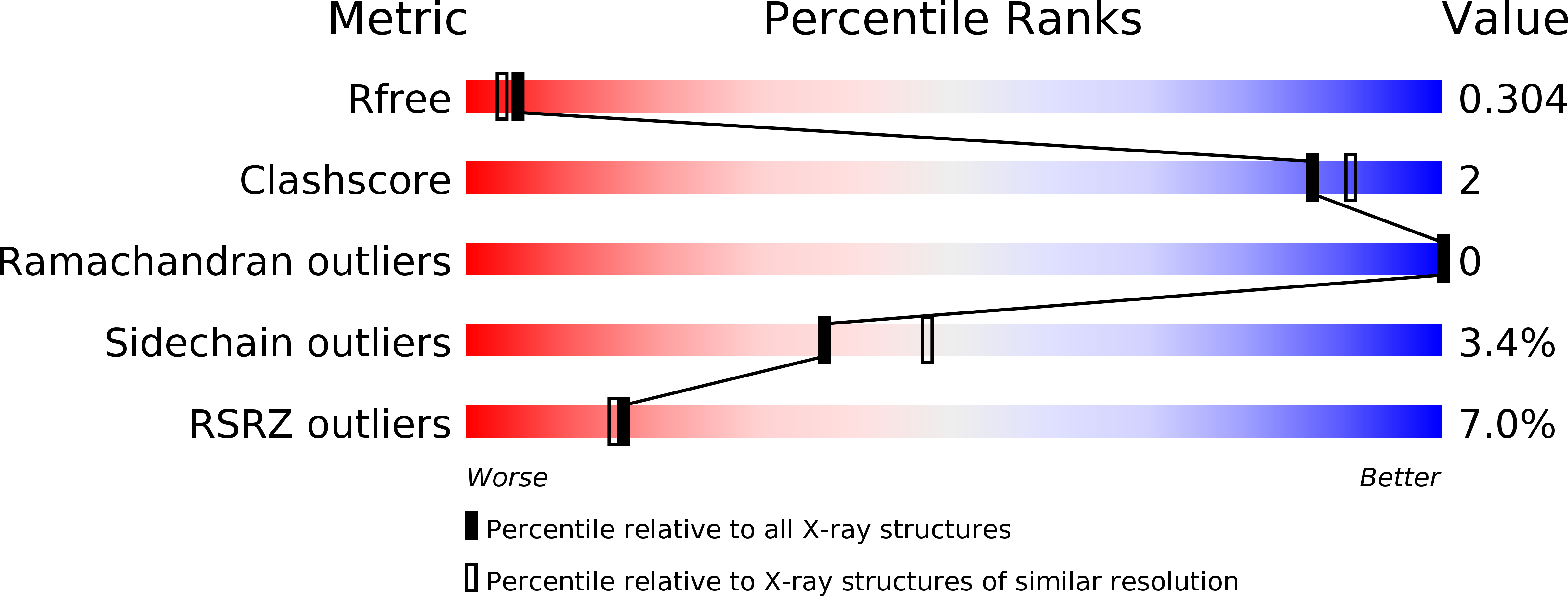
Resolution:
2.20 Å
R-Value Free:
0.29
R-Value Work:
0.24
R-Value Observed:
0.25
Space Group:
P 43 21 2