Deposition Date
2010-07-27
Release Date
2011-08-31
Last Version Date
2021-10-06
Entry Detail
PDB ID:
3O4Q
Keywords:
Title:
Crystal structure of the Rous Associated Virus Integrase catalytic domain A182T in citrate buffer pH 6.2
Biological Source:
Source Organism(s):
Rous sarcoma virus (Taxon ID: 11886)
Expression System(s):
Method Details:
Experimental Method:
Resolution:
1.55 Å
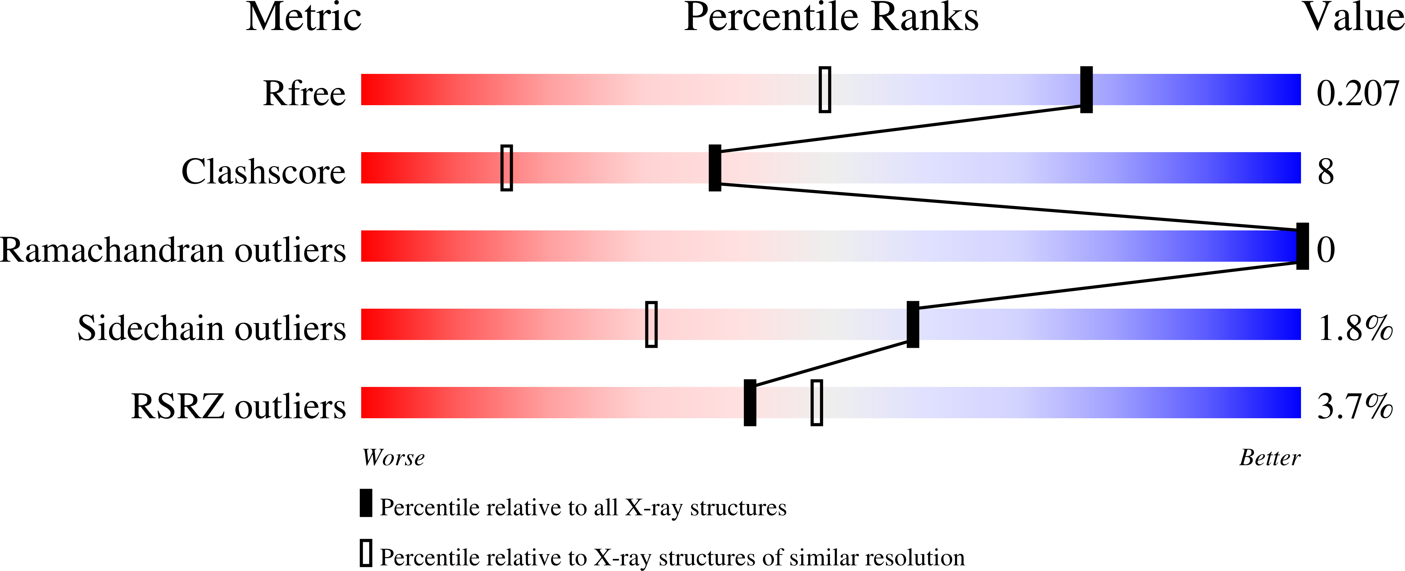
R-Value Free:
0.21
R-Value Work:
0.16
R-Value Observed:
0.16
Space Group:
P 43 21 2