3O48 image
Deposition Date 2010-07-26
Release Date 2011-08-10
Last Version Date 2023-09-06
Entry Detail
PDB ID:
3O48
Keywords:
Title:
Crystal structure of fission protein Fis1 from Saccharomyces cerevisiae
Biological Source:
Source Organism:
Host Organism:
Method Details:
Experimental Method:
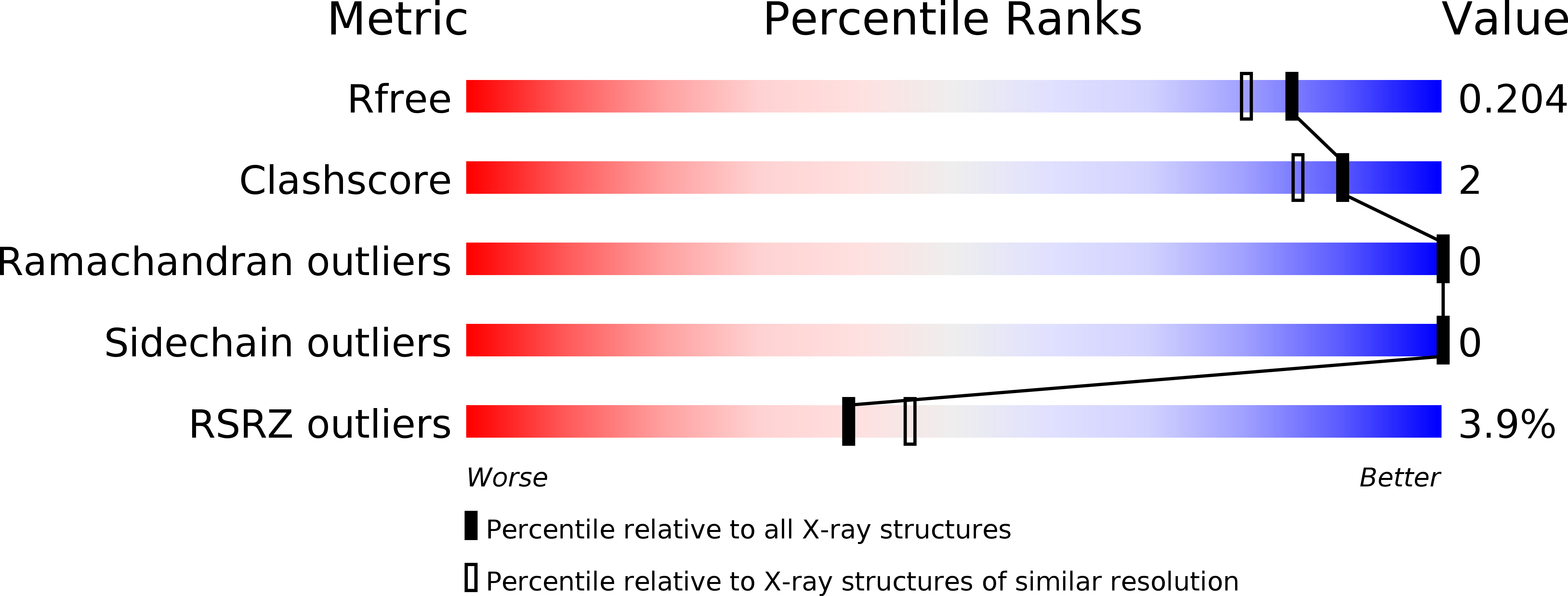
Resolution:
1.75 Å
R-Value Free:
0.18
R-Value Work:
0.16
R-Value Observed:
0.16
Space Group:
P 43 21 2
Macromolecular Entities
Structures with similar UniProt ID
Protein Blast
Polymer Type:polypeptide(L)
Molecule:Mitochondria fission 1 protein
Gene (Uniprot):FIS1
Chain IDs:A
Chain Length:134
Number of Molecules:1
Biological Source:Saccharomyces cerevisiae
Primary Citation
The 1.75 Angstrom resolution structure of fission protein Fis1 from Saccharomyces cerevisiae reveals elusive interactions of the autoinhibitory domain
Acta Crystallogr.,Sect.F 67 1310 1315 (2011)
PMID: 22102223 DOI: 10.1107/S1744309111029368

Abstact

Fis1 mediates mitochondrial and peroxisomal fission. It is tail-anchored to these organelles by a transmembrane domain, exposing a soluble cytoplasmic domain. Previous studies suggested that Fis1 is autoinhibited by its N-terminal region. Here, a 1.75 Å resolution crystal structure of the Fis1 cytoplasmic domain from Saccharomyces cerevisiae is reported which adopts a tetratricopeptide-repeat fold. It is observed that this fold creates a concave surface important for fission, but is sterically occluded by its N-terminal region. Thus, this structure provides a physical basis for autoinhibition and allows a detailed examination of the interactions that stabilize the inhibited state of this molecule.

Legend

Protein

Chemical

Disease

Primary Citation of related structures