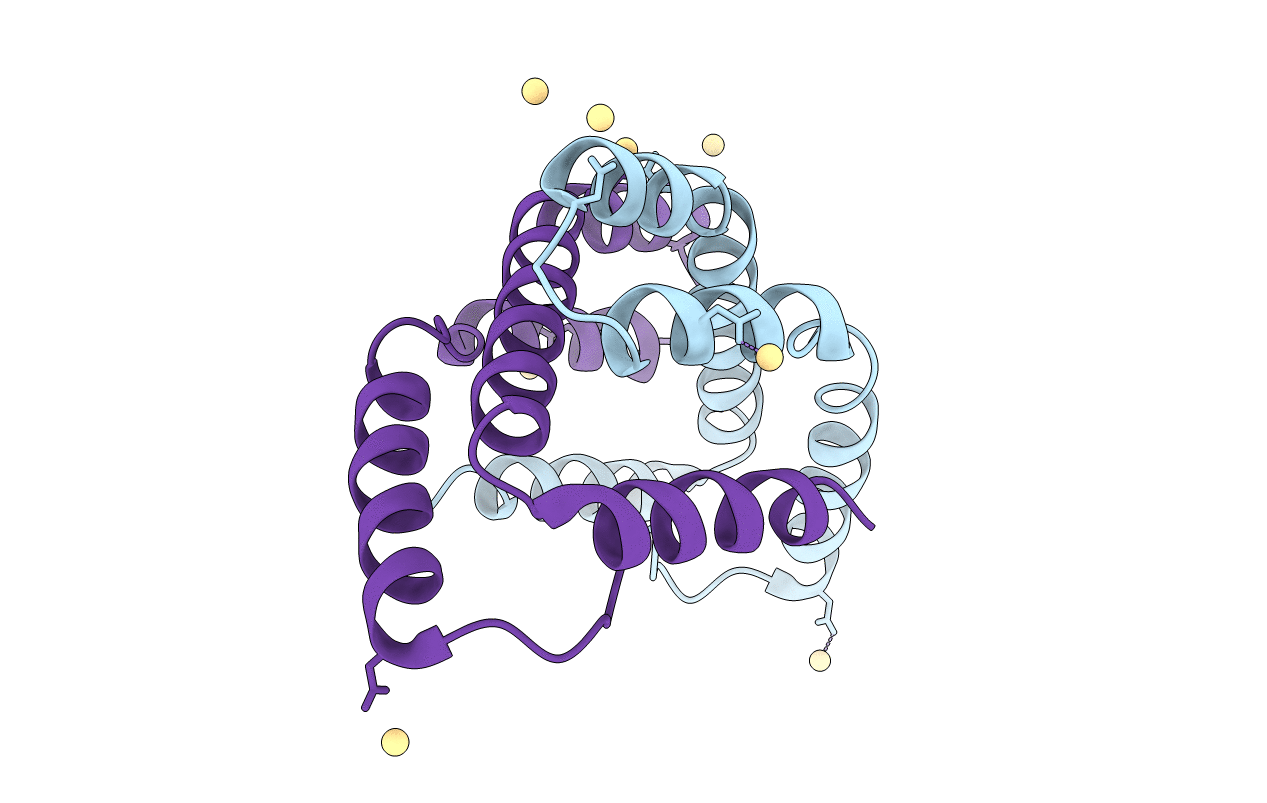
3O39 image
Entry Detail
PDB ID:
3O39
Keywords:
Title:
Crystal Structure of SPY
Biological Source:
Source Organism:
Host Organism:
PDB Version:
Deposition Date:
2010-07-23
Release Date:
2011-02-16
Method Details:
Experimental Method:
Resolution:
2.60 Å
R-Value Free:
0.28
R-Value Work:
0.24
R-Value Observed:
0.24
Space Group:
P 62
Macromolecular Entities
Polymer Type:polypeptide(L)
Description:Periplasmic protein related to spheroblast formation
Chain IDs:A, B
Chain Length:108
Number of Molecules:2
Biological Source:Escherichia coli
Modified Residue
Compound ID Chain ID Parent Comp ID Details 2D Image
MSE A MET SELENOMETHIONINE
Ligand Molecules
Primary Citation
Genetic selection designed to stabilize proteins uncovers a chaperone called Spy.
Nat.Struct.Mol.Biol. 18 262 269 (2011)
PMID: 21317898 DOI: 10.1038/nsmb.2016

Abstact

To optimize the in vivo folding of proteins, we linked protein stability to antibiotic resistance, thereby forcing bacteria to effectively fold and stabilize proteins. When we challenged Escherichia coli to stabilize a very unstable periplasmic protein, it massively overproduced a periplasmic protein called Spy, which increases the steady-state levels of a set of unstable protein mutants up to 700-fold. In vitro studies demonstrate that the Spy protein is an effective ATP-independent chaperone that suppresses protein aggregation and aids protein refolding. Our strategy opens up new routes for chaperone discovery and the custom tailoring of the in vivo folding environment. Spy forms thin, apparently flexible cradle-shaped dimers. The structure of Spy is unlike that of any previously solved chaperone, making it the prototypical member of a new class of small chaperones that facilitate protein refolding in the absence of energy cofactors.

Legend

Protein

Chemical

Disease

Primary Citation of related structures