Deposition Date
2010-07-22
Release Date
2010-09-15
Last Version Date
2024-02-21
Entry Detail
PDB ID:
3O2G
Keywords:
Title:
Crystal Structure of Human gamma-butyrobetaine,2-oxoglutarate dioxygenase 1 (BBOX1)
Biological Source:
Source Organism(s):
Homo sapiens (Taxon ID: 9606)
Expression System(s):
Method Details:
Experimental Method:
Resolution:
1.78 Å
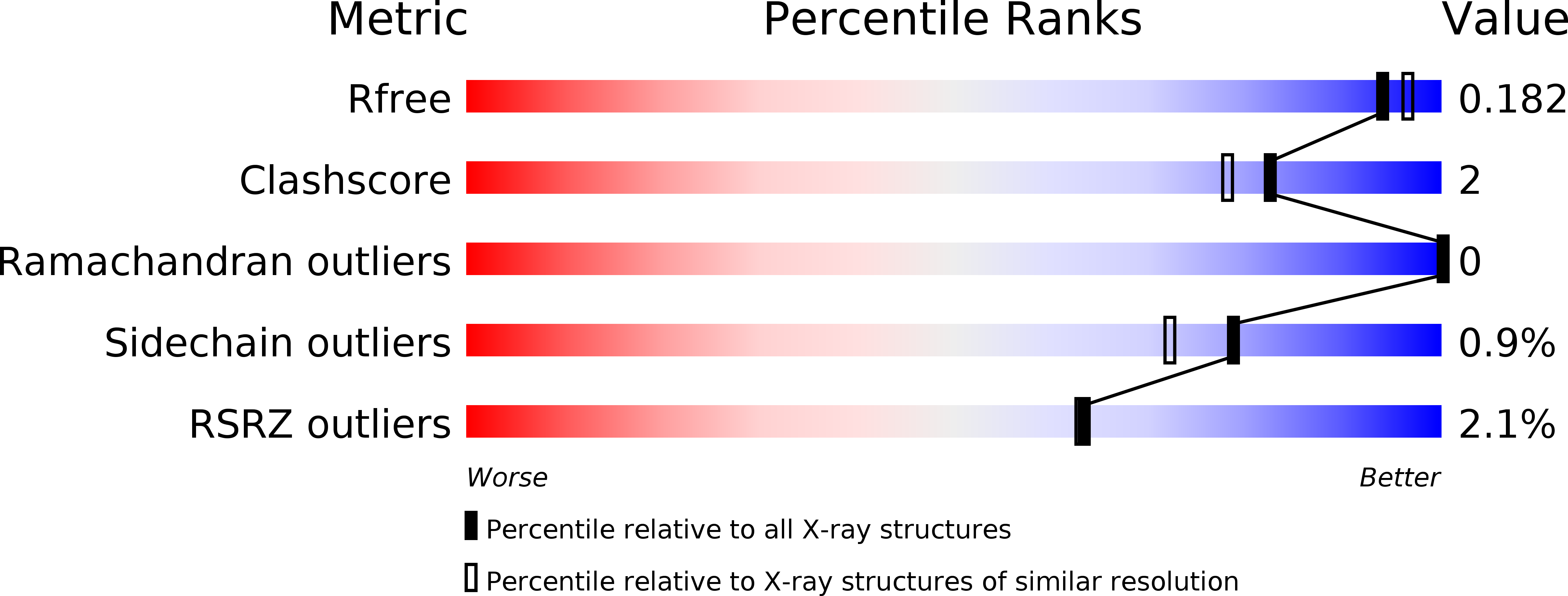
R-Value Free:
0.17
R-Value Work:
0.14
R-Value Observed:
0.14
Space Group:
H 3 2