3O1Q image
Deposition Date 2010-07-21
Release Date 2010-08-18
Last Version Date 2024-02-21
Entry Detail
PDB ID:
3O1Q
Title:
Native Crystal Structure of Helicobacter pylori Urease Accessory Protein UreF
Biological Source:
Source Organism:
Host Organism:
Method Details:
Experimental Method:
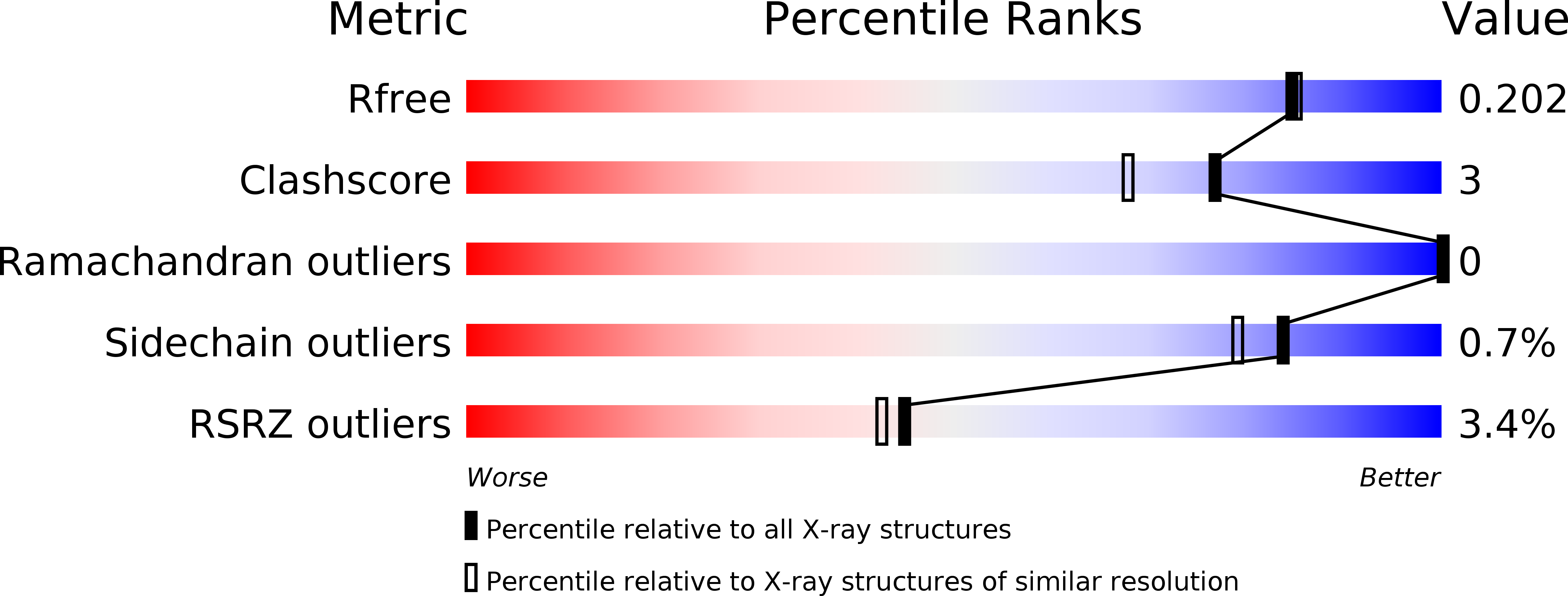
Resolution:
1.85 Å
R-Value Free:
0.20
R-Value Work:
0.16
R-Value Observed:
0.16
Space Group:
C 1 2 1
Macromolecular Entities
Polymer Type:polypeptide(L)
Molecule:Urease accessory protein ureF
Gene (Uniprot):ureF
Chain IDs:A, B, C
Chain Length:254
Number of Molecules:3
Biological Source:Helicobacter pylori
Primary Citation
Assembly of Preactivation Complex for Urease Maturation in Helicobacter pylori: CRYSTAL STRUCTURE OF UreF-UreH PROTEIN COMPLEX.
J.Biol.Chem. 286 43241 43249 (2011)
PMID: 22013070 DOI: 10.1074/jbc.M111.296830

Abstact

Colonization of Helicobacter pylori in the acidic environment of the human stomach depends on the neutralizing activity of urease. Activation of apo-urease involves carboxylation of lysine 219 and insertion of two nickel ions. In H. pylori, this maturation process involves four urease accessory proteins as follows: UreE, UreF, UreG, and UreH. It is postulated that the apo-urease interacts with UreF, UreG, and UreH to form a pre-activation complex that undergoes GTP-dependent activation of urease. The crystal structure of the UreF-UreH complex reveals conformational changes in two distinct regions of UreF upon complex formation. First, the flexible C-terminal residues of UreF become ordered, forming an extra helix α10 and a loop structure stabilized by hydrogen bonds involving Arg-250. Second, the first turn of helix α2 uncoils to expose a conserved residue, Tyr-48. Substitution of R250A or Y48A in UreF abolishes the formation of the heterotrimeric complex of UreG-UreF-UreH and abolishes urease maturation. Our results suggest that the C-terminal residues and helix α2 of UreF are essential for the recruitment of UreG for the formation of the pre-activation complex. The molecular mass of the UreF-UreH complex determined by static light scattering was 116 ± 2.3 kDa, which is consistent with the quaternary structure of a dimer of heterodimers observed in the crystal structure. Taking advantage of the unique 2-fold symmetry observed in both the crystal structures of H. pylori urease and the UreF-UreH complex, we proposed a topology model of the pre-activation complex for urease maturation.

Legend

Protein

Chemical

Disease

Primary Citation of related structures