
Deposition Date
2010-07-19
Release Date
2010-12-08
Last Version Date
2023-09-20
Entry Detail
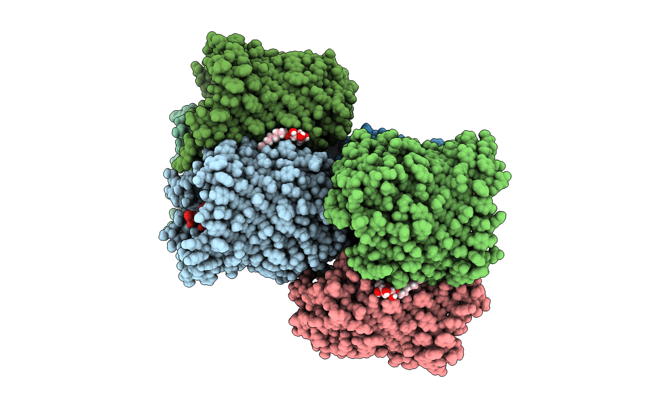
PDB ID:
3O0E
Keywords:
Title:
Crystal structure of OmpF in complex with colicin peptide OBS1
Biological Source:
Source Organism(s):
Escherichia coli (Taxon ID: 562)
Method Details:
Experimental Method:
Resolution:
3.01 Å
R-Value Free:
0.30
R-Value Work:
0.26
R-Value Observed:
0.26
Space Group:
P 1 21 1
