3O07 image
Entry Detail
PDB ID:
3O07
Title:
Crystal structure of yeast pyridoxal 5-phosphate synthase Snz1 complexed with substrate G3P
Biological Source:
Source Organism:
Host Organism:
PDB Version:
Deposition Date:
2010-07-19
Release Date:
2010-11-24
Method Details:
Experimental Method:
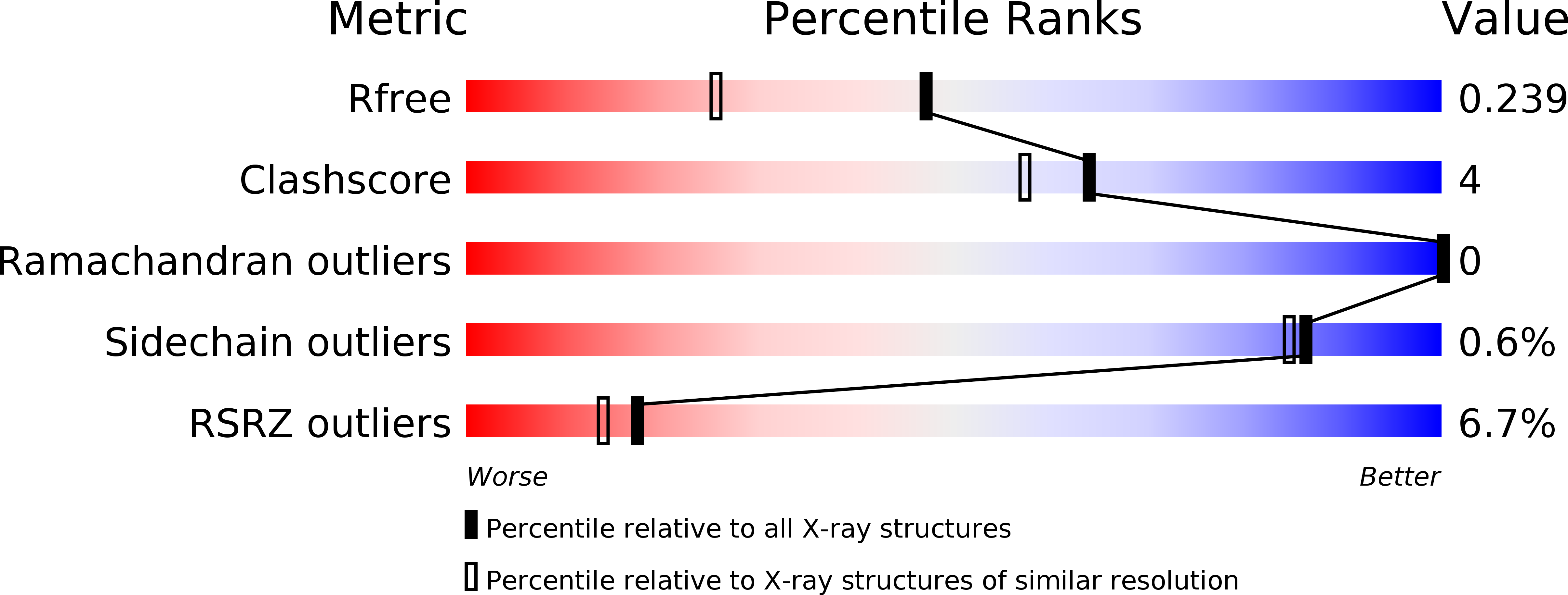
Resolution:
1.80 Å
R-Value Free:
0.23
R-Value Work:
0.20
R-Value Observed:
0.20
Space Group:
P 2 21 21
Macromolecular Entities
Polymer Type:polypeptide(L)
Description:Pyridoxine biosynthesis protein SNZ1
Chain IDs:A, B, C
Chain Length:291
Number of Molecules:3
Biological Source:Saccharomyces cerevisiae
Ligand Molecules
Primary Citation
Structural insights into the catalytic mechanism of the yeast pyridoxal 5-phosphate synthase Snz1
Biochem.J. 432 445 450 (2010)
PMID: 20919991 DOI: 10.1042/BJ20101241

Abstact

In most eubacteria, fungi, apicomplexa, plants and some metazoans, the active form of vitamin B6, PLP (pyridoxal 5-phosphate), is de novo synthesized from three substrates, R5P (ribose 5-phosphate), DHAP (dihydroxyacetone phosphate) and ammonia hydrolysed from glutamine by a complexed glutaminase. Of the three active sites of DXP (deoxyxylulose 5-phosphate)independent PLP synthase (Pdx1), the R5P isomerization site has been assigned, but the sites for DHAP isomerization and PLP formation remain unknown. In the present study, we present the crystal structures of yeast Pdx1/Snz1, in apo-, G3P (glyceraldehyde 3-phosphate)- and PLP-bound forms, at 2.3, 1.8 and 2.2 Å (1 Å=0.1 nm) respectively. Structural and biochemical analysis enabled us to assign the PLP-formation site, a G3P-binding site and a G3P-transfer site. We propose a putative catalytic mechanism for Pdx1/Snz1 in which R5P and DHAP are isomerized at two distinct sites and transferred along well-defined routes to a final destination for PLP synthesis.

Legend

Protein

Chemical

Disease

Primary Citation of related structures