Abstact
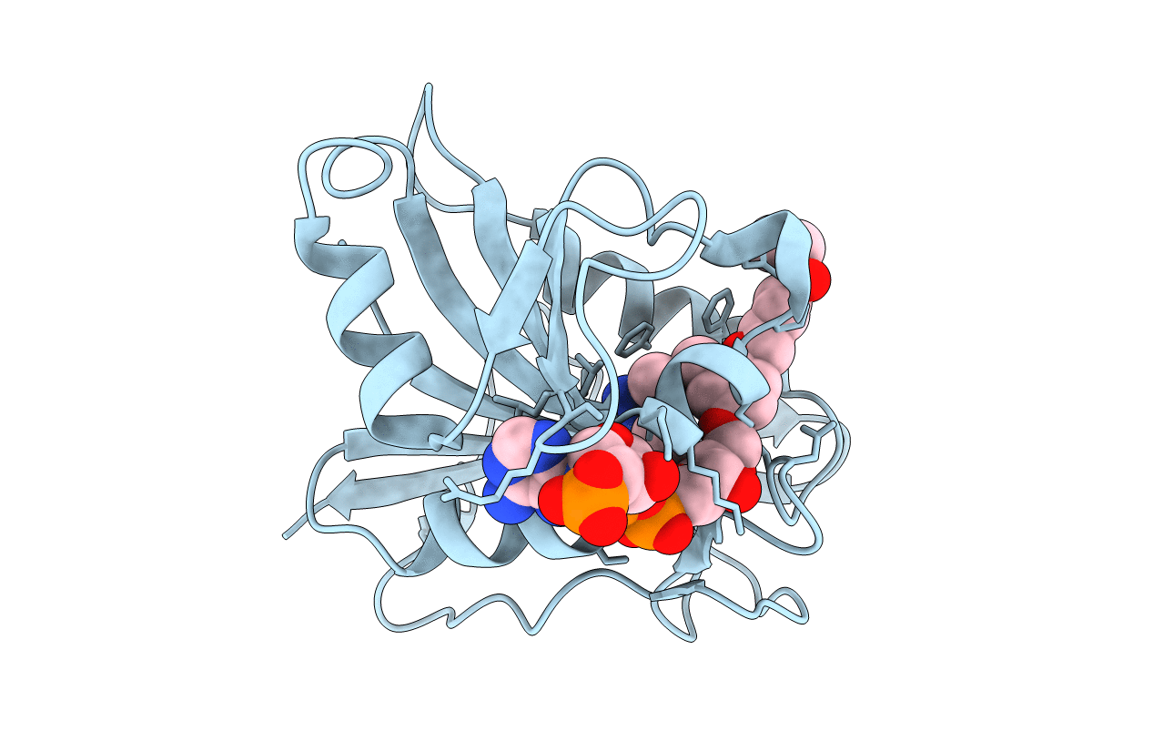
Structural data are reported for five antifolates, namely 2,4-diamino-6-[5'-(5-carboxypentyloxy)-2'-methoxybenzyl]-5-methylpyrido[2,3-d]pyrimidine, (1), and the 5'-[3-(ethoxycarbonyl)propoxy]-, (2), 5'-[3-(ethoxycarbonyl)butoxy]-, (3), 5'-[3-(ethoxycarbonyl)pentyloxy]-, (4), and 5'-benzyloxy-, (5), derivatives, which are potent and selective for Pneumocystis carinii dihydrofolate reductase (pcDHFR). Crystal structures are reported for their ternary complexes with NADPH and pcDHFR refined to between 1.4 and 2.0 Å resolution and for that of 3 with human DHFR (hDHFR) to 1.8 Å resolution. These data reveal that the carboxylate of the ω-carboxyalkoxy side chain of 1, the most potent inhibitor in this series, forms ionic interactions with the conserved Arg75 in the substrate-binding pocket of pcDHFR, whereas the less potent ethyl esters of 2-4 bind with variable side-chain conformations. The benzyloxy side chain of 5 makes no contact with Arg75 and is the least active inhibitor in this series. These structural results suggest that the weaker binding of this series compared with that of their pyrimidine homologs in part arises from the flexibility observed in their side-chain conformations, which do not optimize intermolecular contact to Arg75. Structural data for the binding of 3 to both hDHFR and pcDHFR reveals that the inhibitor binds in two different conformations, one similar to each of the two conformations observed for the parent pyrido[2,3-d]pyrimidine, piritrexim (PTX), bound to hDHFR. The structure of the pcDHFR complex of 4 reveals disorder in the side-chain orientation; one orientation has the ω-carboxyalkoxy side chain positioned in the folate-binding pocket similar to the others in this series, while the second orientation occupies a new site near the nicotinamide ring of NADPH. This alternate binding site has not been observed in other DHFR structures. Structural data for the pcDHFR complex of 5 show that its benzyl side chain forms intermolecular van der Waals interactions with Phe69 in the binding pocket that could account for its enhanced binding selectivity compared with the other analogs in this series.