Deposition Date
2010-07-15
Release Date
2010-09-22
Last Version Date
2024-10-16
Entry Detail
PDB ID:
3NYO
Keywords:
Title:
Crystal Structure of G Protein-Coupled Receptor Kinase 6 in Complex with AMP
Biological Source:
Source Organism(s):
Homo sapiens (Taxon ID: 9606)
Expression System(s):
Method Details:
Experimental Method:
Resolution:
2.92 Å
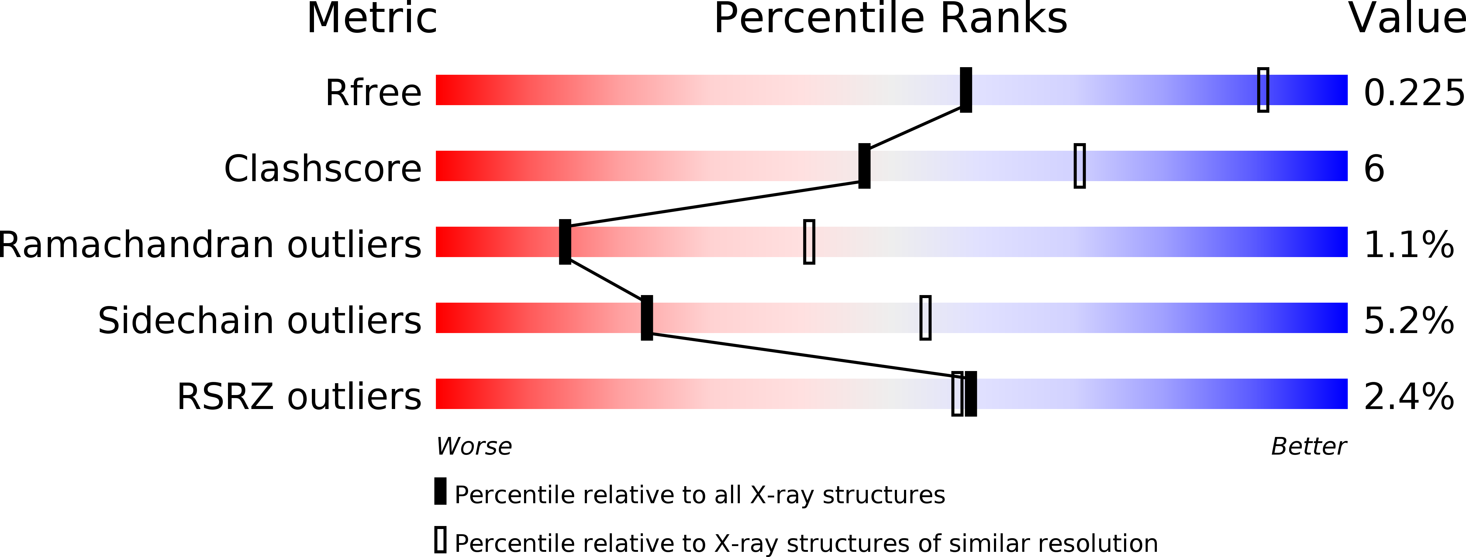
R-Value Free:
0.25
R-Value Work:
0.22
R-Value Observed:
0.22
Space Group:
P 61