Deposition Date
2010-07-15
Release Date
2010-10-20
Last Version Date
2024-02-21
Entry Detail
PDB ID:
3NYK
Keywords:
Title:
The structure of cobalt-substituted pseudoazurin from Alcaligenes faecalis
Biological Source:
Source Organism(s):
Alcaligenes faecalis (Taxon ID: 511)
Expression System(s):
Method Details:
Experimental Method:
Resolution:
1.86 Å
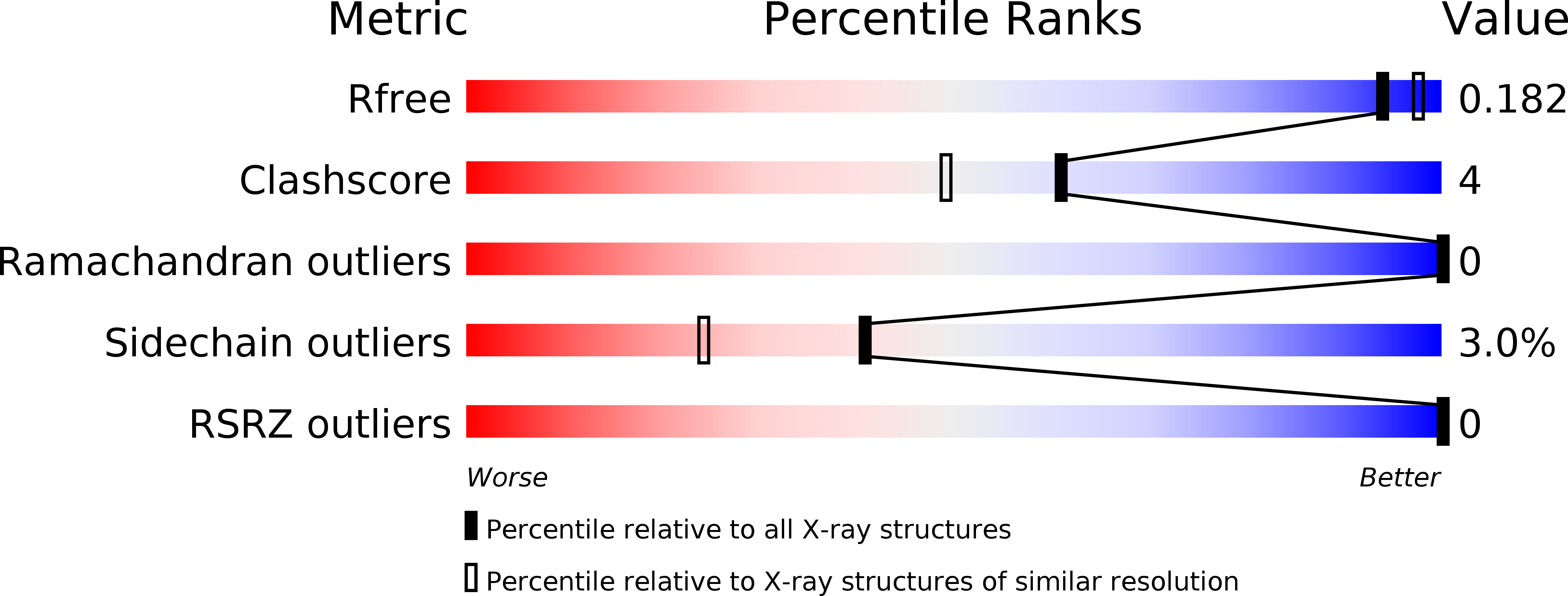
R-Value Free:
0.17
R-Value Work:
0.14
R-Value Observed:
0.15
Space Group:
P 65