3NXZ image
Deposition Date 2010-07-14
Release Date 2010-08-25
Last Version Date 2023-09-06
Entry Detail
PDB ID:
3NXZ
Title:
Crystal Structure of UreE from Helicobacter pylori (Cu2+ bound form)
Biological Source:
Source Organism:
Host Organism:
Method Details:
Experimental Method:
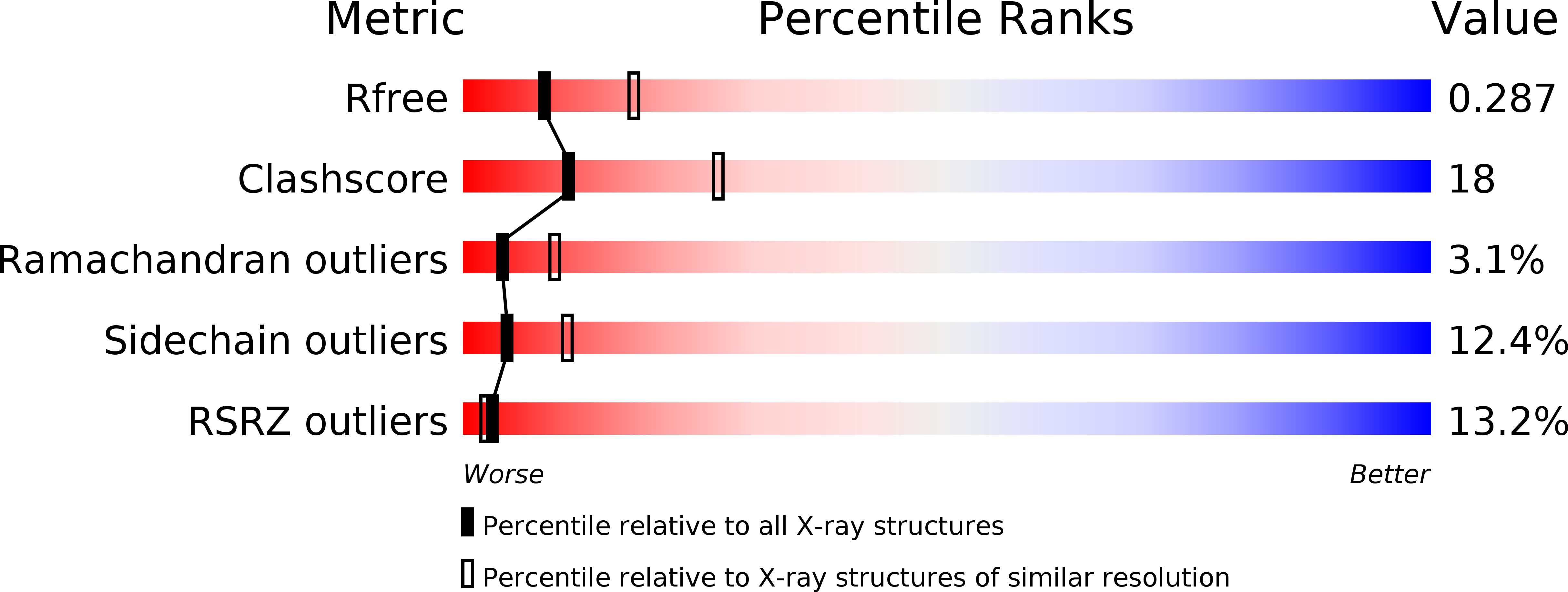
Resolution:
2.70 Å
R-Value Free:
0.29
R-Value Work:
0.25
R-Value Observed:
0.25
Space Group:
P 41 21 2
Macromolecular Entities
Polymer Type:polypeptide(L)
Molecule:Urease accessory protein ureE
Gene (Uniprot):ureE
Chain IDs:A, B, C, D
Chain Length:170
Number of Molecules:4
Biological Source:Helicobacter pylori
Ligand Molecules
Primary Citation
Crystal Structures of Apo and Metal-Bound Forms of the UreE Protein from Helicobacter pylori: Role of Multiple Metal Binding Sites
Biochemistry 49 7080 7088 (2010)
PMID: 20681615 DOI: 10.1021/bi100372h

Abstact

The crystal structure of the urease maturation protein UreE from Helicobacter pylori has been determined in its apo form at 2.1 A resolution, bound to Cu(2+) at 2.7 A resolution, and bound to Ni(2+) at 3.1 A resolution. Apo UreE forms dimers, while the metal-bound enzymes are arranged as tetramers that consist of a dimer of dimers associated around the metal ion through coordination by His102 residues from each subunit of the tetramer. Comparison of independent subunits from different crystal forms indicates changes in the relative arrangement of the N- and C-terminal domains in response to metal binding. The improved ability of engineered versions of UreE containing hexahistidine sequences at either the N-terminal or C-terminal end to provide Ni(2+) for the final metal sink (urease) is eliminated in the H102A version. Therefore, the ability of the improved Ni(2+)-binding versions to deliver more nickel is likely an effect of an increased local concentration of metal ions that can rapidly replenish transferred ions bound to His102.

Legend

Protein

Chemical

Disease

Primary Citation of related structures