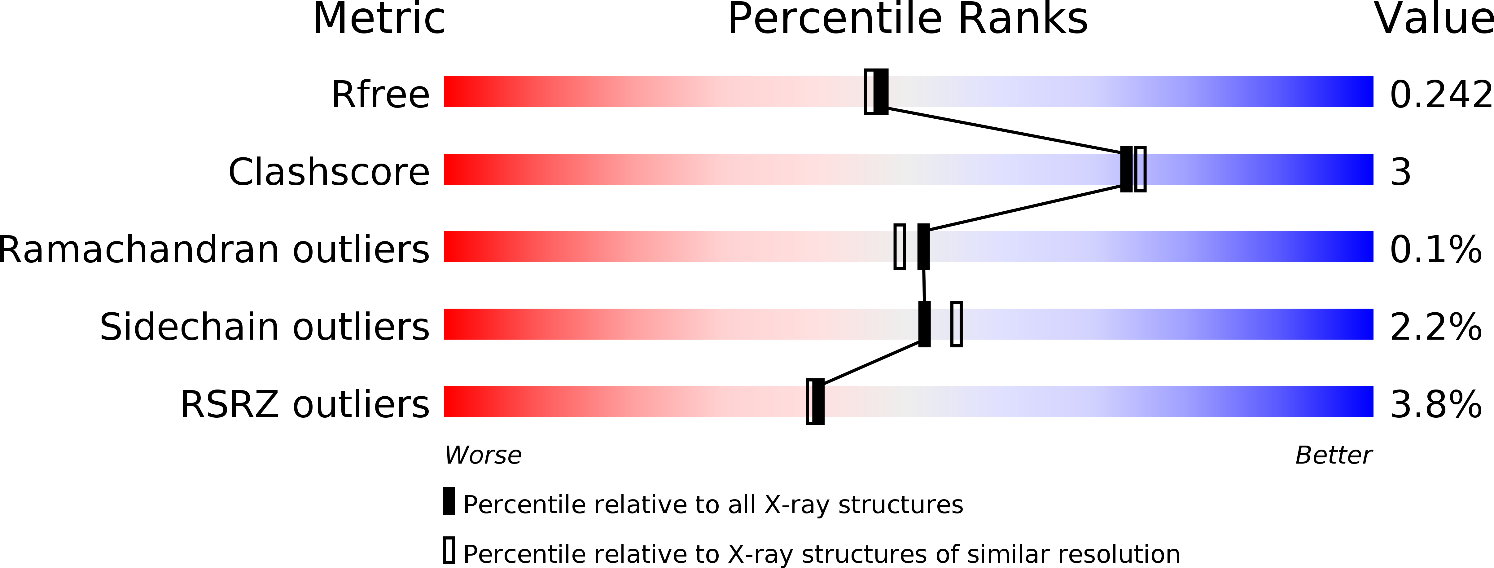
3NXQ image
Deposition Date 2010-07-14
Release Date 2010-09-08
Last Version Date 2024-10-16
Entry Detail
PDB ID:
3NXQ
Title:
Angiotensin Converting Enzyme N domain glycsoylation mutant (Ndom389) in complex with RXP407
Biological Source:
Source Organism:
Homo sapiens (Taxon ID: 9606)
Host Organism:
Method Details:
Experimental Method:
Resolution:
1.99 Å
R-Value Free:
0.23
R-Value Work:
0.19
R-Value Observed:
0.19
Space Group:
P 1
Macromolecular Entities
Polymer Type:polypeptide(L)
Molecule:Angiotensin-converting enzyme
Gene (Uniprot):ACE
Mutations:N9Q, N25Q, N82Q, N117Q, N131Q, N289Q, Q545R, P576L, R629L
Chain IDs:A, B
Chain Length:629
Number of Molecules:2
Biological Source:Homo sapiens
Modified Residue
Compound ID Chain ID Parent Comp ID Details 2D Image
ASN A ASN GLYCOSYLATION SITE
Peptide-like Molecules
PRD_001050
Primary Citation
The N domain of human angiotensin-I-converting enzyme: the role of N-glycosylation and the crystal structure in complex with an N domain-specific phosphinic inhibitor, RXP407.
J.Biol.Chem. 285 35685 35693 (2010)
PMID: 20826823 DOI: 10.1074/jbc.M110.167866

Abstact

Angiotensin-I-converting enzyme (ACE) plays a critical role in the regulation of blood pressure through its central role in the renin-angiotensin and kallikrein-kinin systems. ACE contains two domains, the N and C domains, both of which are heavily glycosylated. Structural studies of ACE have been fraught with severe difficulties because of surface glycosylation of the protein. In order to investigate the role of glycosylation in the N domain and to create suitable forms for crystallization, we have investigated the importance of the 10 potential N-linked glycan sites using enzymatic deglycosylation, limited proteolysis, and mass spectrometry. A number of glycosylation mutants were generated via site-directed mutagenesis, expressed in CHO cells, and analyzed for enzymatic activity and thermal stability. At least eight of 10 of the potential glycan sites are glycosylated; three C-terminal sites were sufficient for expression of active N domain, whereas two N-terminal sites are important for its thermal stability. The minimally glycosylated Ndom389 construct was highly suitable for crystallization studies. The structure in the presence of an N domain-selective phosphinic inhibitor RXP407 was determined to 2.0 Å resolution. The Ndom389 structure revealed a hinge region that may contribute to the breathing motion proposed for substrate binding.

Legend

Protein

Chemical

Disease

Primary Citation of related structures