
Deposition Date
2010-07-12
Release Date
2011-02-16
Last Version Date
2023-11-01
Entry Detail
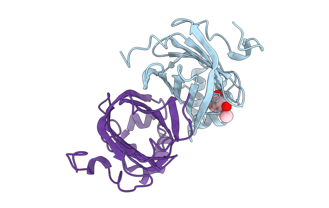
PDB ID:
3NX2
Keywords:
Title:
Enterobacter sp. Px6-4 Ferulic Acid Decarboxylase in complex with substrate analogues
Biological Source:
Source Organism(s):
Enterobacter sp. Px6-4 (Taxon ID: 418698)
Expression System(s):
Method Details:
Experimental Method:
Resolution:
2.01 Å
R-Value Free:
0.23
R-Value Work:
0.18
R-Value Observed:
0.19
Space Group:
P 1 21 1
