Deposition Date
2010-07-09
Release Date
2010-08-04
Last Version Date
2023-11-15
Entry Detail
PDB ID:
3NW3
Keywords:
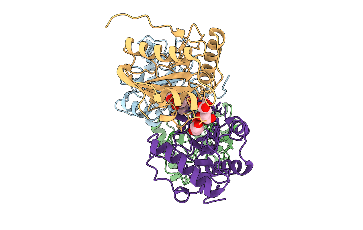
Title:
Crystal structure of the complex of peptidoglycan recognition protein (PGRP-S) with the PGN Fragment at 2.5 A resolution
Biological Source:
Source Organism(s):
Camelus dromedarius (Taxon ID: 9838)
Method Details:
Experimental Method:
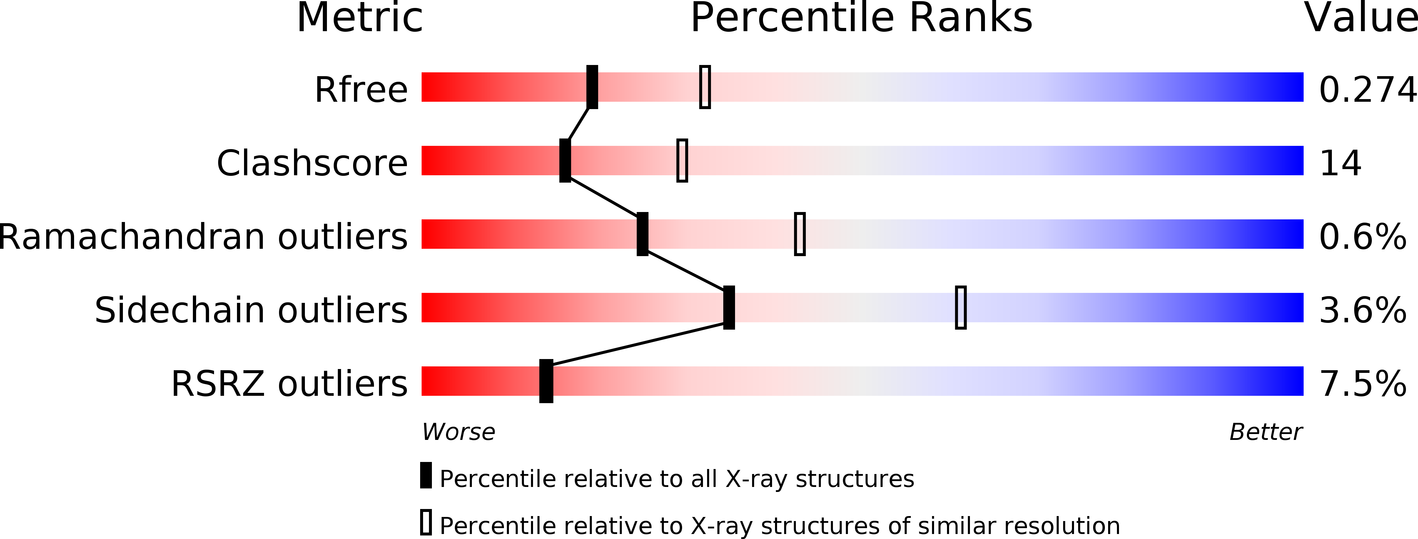
Resolution:
2.50 Å
R-Value Free:
0.24
R-Value Work:
0.23
R-Value Observed:
0.23
Space Group:
I 2 2 2