3NVX image
Entry Detail
PDB ID:
3NVX
Keywords:
Title:
Molecular mechanism of guidance cue recognition
Biological Source:
Source Organism:
Host Organism:
PDB Version:
Deposition Date:
2010-07-08
Release Date:
2010-09-15
Method Details:
Experimental Method:
Resolution:
2.00 Å
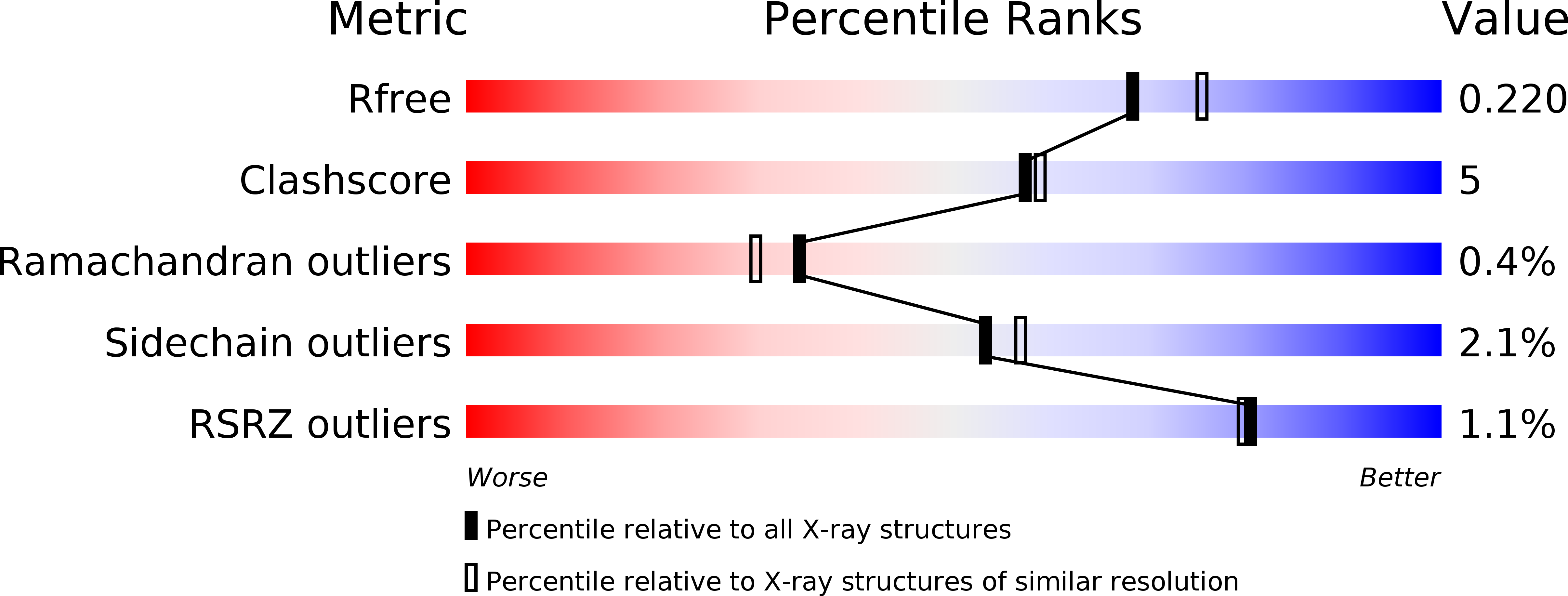
R-Value Free:
0.22
R-Value Work:
0.17
R-Value Observed:
0.17
Space Group:
C 2 2 21
Macromolecular Entities
Polymer Type:polypeptide(L)
Description:Protein A39
Chain IDs:A, B
Chain Length:383
Number of Molecules:2
Biological Source:Vaccinia virus
Modified Residue
Compound ID Chain ID Parent Comp ID Details 2D Image
ASN B ASN GLYCOSYLATION SITE
Ligand Molecules
Primary Citation
Structural Basis of Semaphorin-Plexin Recognition and Viral Mimicry from Sema7A and A39R Complexes with PlexinC1.
Cell(Cambridge,Mass.) 142 749 761 (2010)
PMID: 20727575 DOI: 10.1016/j.cell.2010.07.040

Abstact

Repulsive signaling by Semaphorins and Plexins is crucial for the development and homeostasis of the nervous, immune, and cardiovascular systems. Sema7A acts as both an immune and a neural Semaphorin through PlexinC1, and A39R is a Sema7A mimic secreted by smallpox virus. We report the structures of Sema7A and A39R complexed with the Semaphorin-binding module of PlexinC1. Both structures show two PlexinC1 molecules symmetrically bridged by Semaphorin dimers, in which the Semaphorin and PlexinC1 beta propellers interact in an edge-on, orthogonal orientation. Both binding interfaces are dominated by the insertion of the Semaphorin's 4c-4d loop into a deep groove in blade 3 of the PlexinC1 propeller. A39R appears to achieve Sema7A mimicry by preserving key Plexin-binding determinants seen in the mammalian Sema7A complex that have evolved to achieve higher affinity binding to the host-derived PlexinC1. The complex structures support a conserved Semaphorin-Plexin recognition mode and suggest that Plexins are activated by dimerization.

Legend

Protein

Chemical

Disease

Primary Citation of related structures