Deposition Date
2010-07-08
Release Date
2010-11-03
Last Version Date
2023-11-01
Entry Detail
Biological Source:
Source Organism(s):
Novosphingobium aromaticivorans (Taxon ID: 279238)
Expression System(s):
Method Details:
Experimental Method:
Resolution:
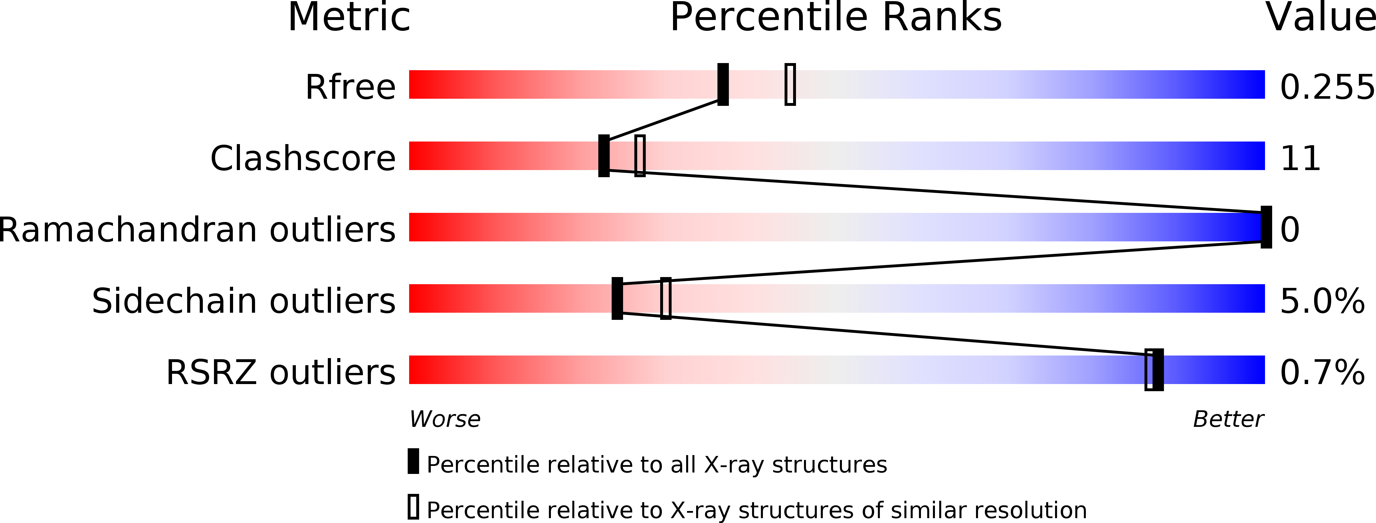
2.20 Å
R-Value Free:
0.25
R-Value Work:
0.18
R-Value Observed:
0.19
Space Group:
P 32 2 1