3NUJ image
Deposition Date 2010-07-07
Release Date 2010-08-25
Last Version Date 2024-02-21
Entry Detail
PDB ID:
3NUJ
Title:
Crystal Structure of HIV-1 Protease Mutant I54V with Antiviral Drug Amprenavir
Biological Source:
Source Organism:
Method Details:
Experimental Method:
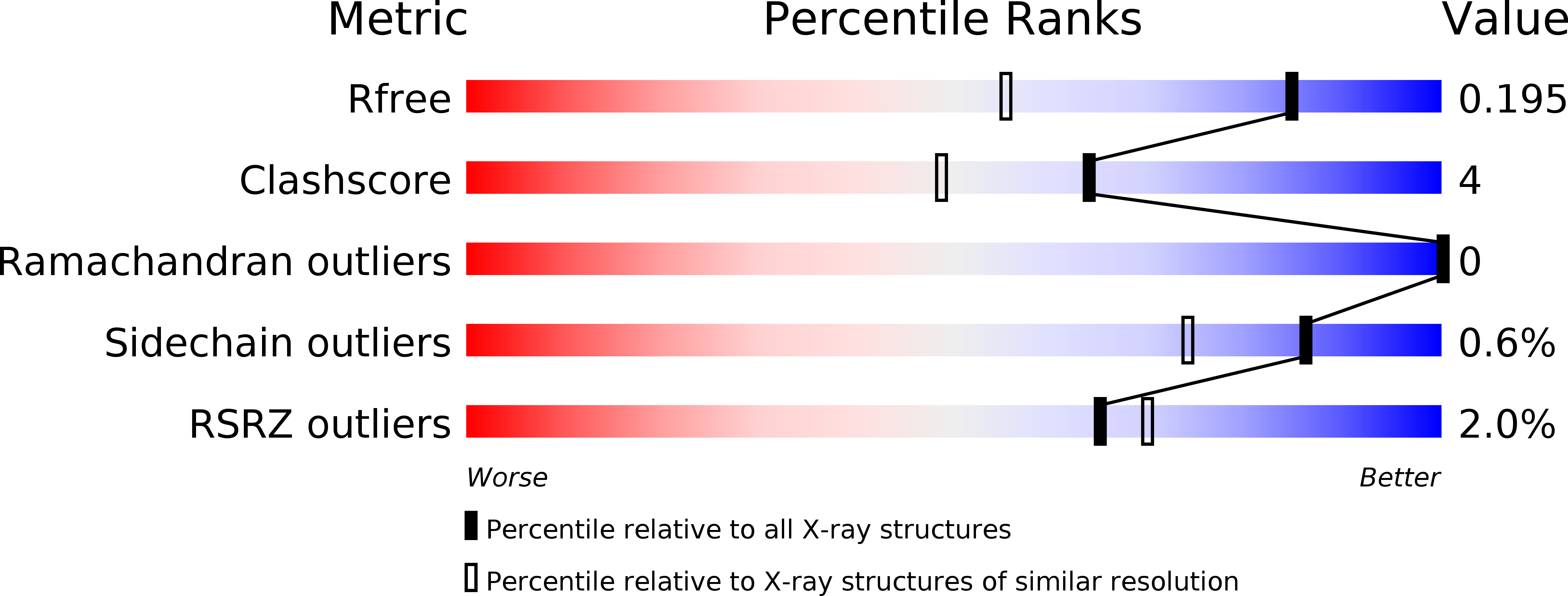
Resolution:
1.50 Å
R-Value Free:
0.19
R-Value Observed:
0.14
Space Group:
P 21 21 2
Macromolecular Entities
Structures with similar UniProt ID
Protein Blast
Polymer Type:polypeptide(L)
Molecule:Protease
Gene (Uniprot):gag-pol
Mutagens:Q7K, L33I, I54V, L63I, C67A, C95A
Chain IDs:A, B
Chain Length:99
Number of Molecules:2
Biological Source:Human immunodeficiency virus 1
Primary Citation
Amprenavir complexes with HIV-1 protease and its drug-resistant mutants altering hydrophobic clusters.
Febs J. 277 3699 3714 (2010)
PMID: 20695887 DOI: 10.1111/j.1742-4658.2010.07771.x

Abstact

The structural and kinetic effects of amprenavir (APV), a clinical HIV protease (PR) inhibitor, were analyzed with wild-type enzyme and mutants with single substitutions of V32I, I50V, I54V, I54M, I84V and L90M that are common in drug resistance. Crystal structures of the APV complexes at resolutions of 1.02-1.85 Å reveal the structural changes due to the mutations. Substitution of the larger side chains in PR(V32I) , PR(I54M) and PR(L90M) resulted in the formation of new hydrophobic contacts with flap residues, residues 79 and 80, and Asp25, respectively. Mutation to smaller side chains eliminated hydrophobic interactions in the PR(I50V) and PR(I54V) structures. The PR(I84V)-APV complex had lost hydrophobic contacts with APV, the PR(V32I)-APV complex showed increased hydrophobic contacts within the hydrophobic cluster and the PR(I50V) complex had weaker polar and hydrophobic interactions with APV. The observed structural changes in PR(I84V)-APV, PR(V32I)-APV and PR(I50V)-APV were related to their reduced inhibition by APV of six-, 10- and 30-fold, respectively, relative to wild-type PR. The APV complexes were compared with the corresponding saquinavir complexes. The PR dimers had distinct rearrangements of the flaps and 80's loops that adapt to the different P1' groups of the inhibitors, while maintaining contacts within the hydrophobic cluster. These small changes in the loops and weak internal interactions produce the different patterns of resistant mutations for the two drugs.

Legend

Protein

Chemical

Disease

Primary Citation of related structures