3NUG image
Deposition Date 2010-07-06
Release Date 2011-04-20
Last Version Date 2023-11-01
Entry Detail
PDB ID:
3NUG
Keywords:
Title:
Crystal structure of wild type tetrameric pyridoxal 4-dehydrogenase from Mesorhizobium loti
Biological Source:
Source Organism:
Host Organism:
Method Details:
Experimental Method:
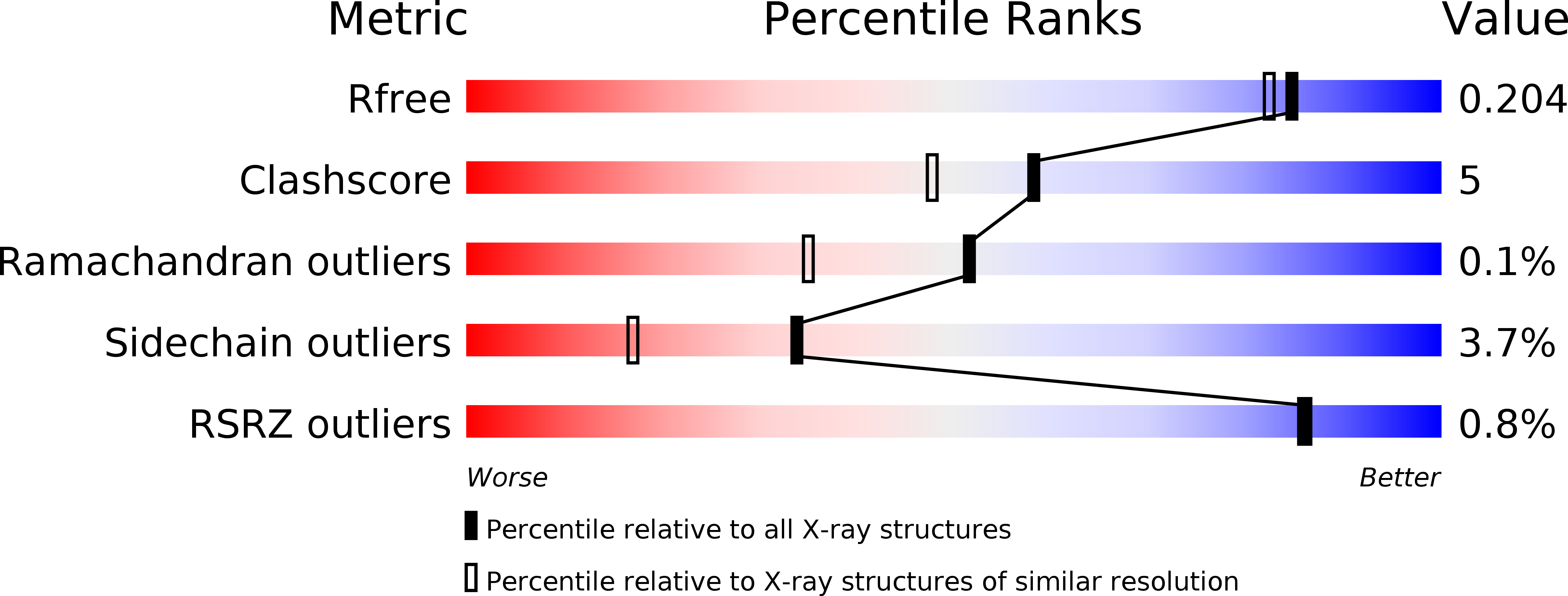
Resolution:
1.79 Å
R-Value Free:
0.20
R-Value Work:
0.15
R-Value Observed:
0.15
Space Group:
P 1 21 1
Macromolecular Entities
Polymer Type:polypeptide(L)
Molecule:3-oxoacyl-(Acyl-carrier protein) reductase
Gene (Uniprot):pldh-t
Chain IDs:A, B, C, D
Chain Length:247
Number of Molecules:4
Biological Source:Mesorhizobium loti
Primary Citation
The crystal structure of SDR-type pyridoxal 4-dehydrogenase of Mesorhizobium loti
Biosci.Biotechnol.Biochem. 75 388 390 (2011)
PMID: 21307574 DOI: 10.1271/bbb.100748

Abstact

Pyridoxal 4-dehydrogenase catalyzes the irreversible oxidation of pyridoxal to 4-pyridoxolactone and is involved in degradation pathway I of pyridoxine, a vitamin B(6) compound. Its crystal structure was elucidated for the first time. Molecular replacement with (S)-1-phenylthanol dehydrogenase (PDB code 2EW8) was adopted to determine the tertiary structure of the NAD(+)-bound enzyme.

Legend

Protein

Chemical

Disease

Primary Citation of related structures