3NU1 image
Deposition Date 2010-07-06
Release Date 2010-10-13
Last Version Date 2024-03-20
Entry Detail
PDB ID:
3NU1
Keywords:
Title:
Structure of holo form of a periplasmic heme binding protein
Biological Source:
Source Organism:
Host Organism:
Method Details:
Experimental Method:
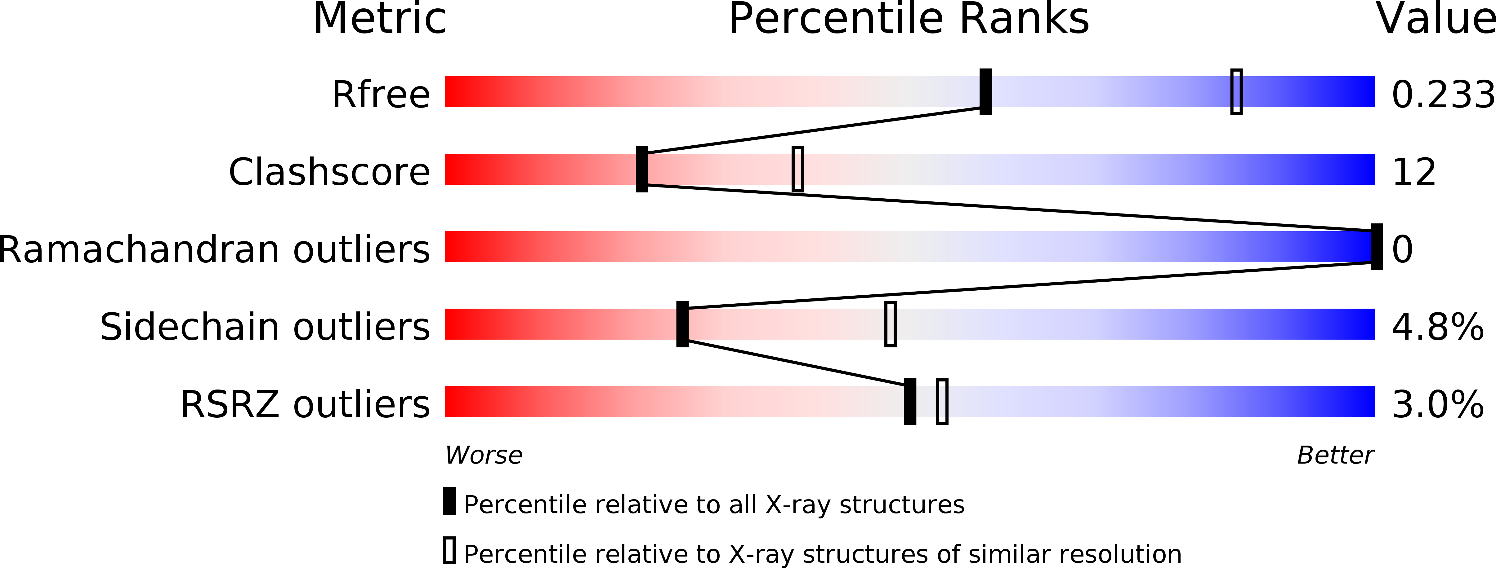
Resolution:
2.50 Å
R-Value Free:
0.23
R-Value Work:
0.19
R-Value Observed:
0.19
Space Group:
P 61 2 2
Macromolecular Entities
Polymer Type:polypeptide(L)
Molecule:Hemin-binding periplasmic protein
Chain IDs:A, B
Chain Length:254
Number of Molecules:2
Biological Source:Yersinia pestis
Ligand Molecules
Primary Citation
Two stacked heme molecules in the binding pocket of the periplasmic heme-binding protein HmuT from Yersinia pestis.
J.Mol.Biol. 404 220 231 (2010)
PMID: 20888343 DOI: 10.1016/j.jmb.2010.09.005

Abstact

The periplasmic binding protein HmuT from Yersinia pestis (YpHmuT) is a component of the heme uptake locus hmu and delivers bound hemin to the inner-membrane-localized, ATP-binding cassette (ABC) transporter HmuUV for translocation into the cytoplasm. The mechanism of this process, heme transport across the inner membrane of pathogenic bacteria, is currently insufficiently understood at the molecular level. Here we describe the crystal structures of the substrate-free and heme-bound states of YpHmuT, revealing two lobes with a central binding cleft. Superposition of the apo and holo states reveals a minor tilting motion of the lobes surrounding concomitant with heme binding. Unexpectedly, YpHmuT binds two stacked hemes in a central binding cleft that is larger than those of the homologous periplasmic heme-binding proteins ShuT and PhuT, both of which bind only one heme. The hemes bound to YpHmuT are coordinated via a tyrosine side chain that contacts the Fe atom of one heme and a histidine that contacts the Fe atom of the other heme. The coordinating histidine is only conserved in a subset of periplasmic heme binding proteins suggesting that its presence predicts the ability to bind two heme molecules simultaneously. The structural data are supported by spectroscopic binding studies performed in solution, where up to two hemes can bind to YpHmuT. Isothermal titration calorimetry suggests that the two hemes are bound in discrete, sequential steps and with dissociation constants (K(D)) of ∼0.29  and ∼29 nM, which is similar to the affinities observed in other bacterial substrate binding proteins. Our findings suggest that the cognate ABC transporter HmuUV may simultaneously translocate two hemes per reaction cycle.

Legend

Protein

Chemical

Disease

Primary Citation of related structures