3NTH image
Entry Detail
PDB ID:
3NTH
Keywords:
Title:
Crystal structure of Tudor and Aubergine [R13(me2s)] complex
Biological Source:
Source Organism:
Host Organism:
PDB Version:
Deposition Date:
2010-07-05
Release Date:
2010-09-15
Method Details:
Experimental Method:
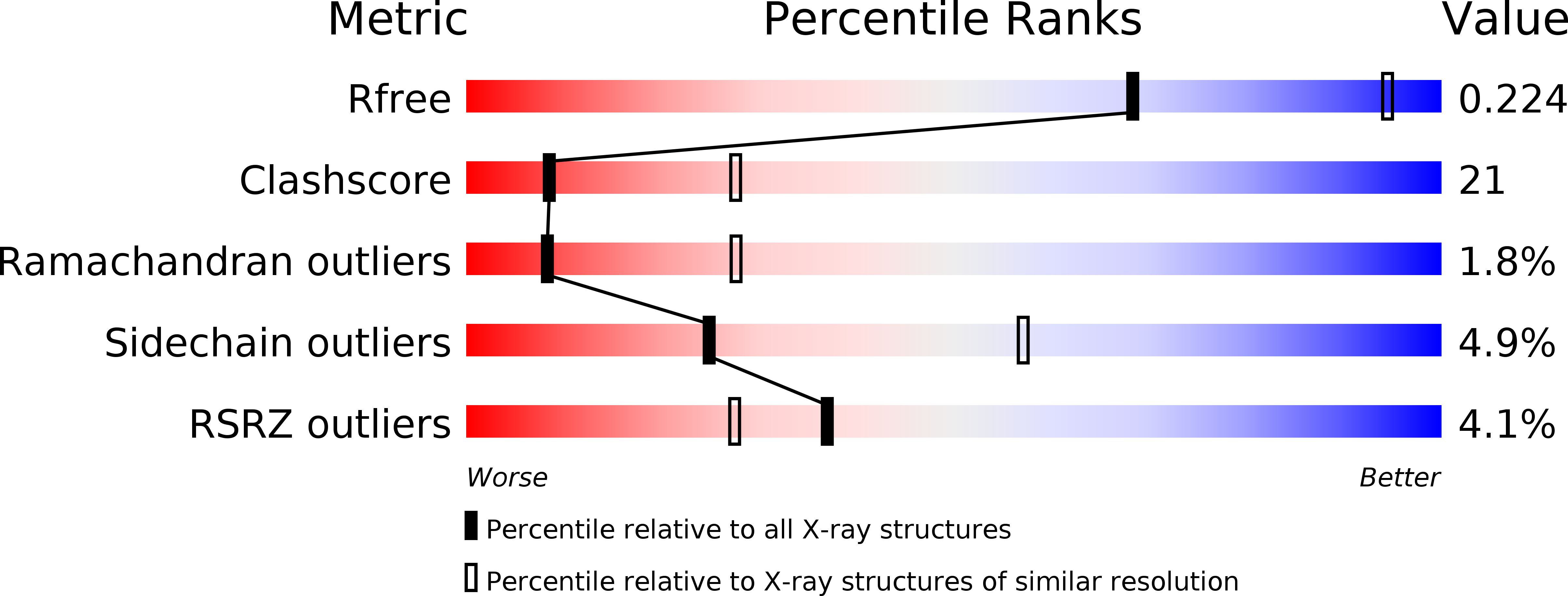
Resolution:
2.80 Å
R-Value Free:
0.27
R-Value Work:
0.21
Space Group:
P 43 21 2
Macromolecular Entities
Polymer Type:polypeptide(L)
Description:Maternal protein tudor
Chain IDs:A
Chain Length:172
Number of Molecules:1
Biological Source:Drosophila melanogaster
Polymer Type:polypeptide(L)
Description:peptide from Aubergine
Chain IDs:B (auth: C)
Chain Length:6
Number of Molecules:1
Biological Source:Drosophila melanogaster
Modified Residue
Compound ID Chain ID Parent Comp ID Details 2D Image
2MR B ARG N3, N4-DIMETHYLARGININE
Primary Citation
Structural basis for methylarginine-dependent recognition of Aubergine by Tudor
Genes Dev. 24 1876 1881 (2010)
PMID: 20713507 DOI: 10.1101/gad.1956010

Abstact

Piwi proteins are modified by symmetric dimethylation of arginine (sDMA), and the methylarginine-dependent interaction with Tudor domain proteins is critical for their functions in germline development. Cocrystal structures of an extended Tudor domain (eTud) of Drosophila Tudor with methylated peptides of Aubergine, a Piwi family protein, reveal that sDMA is recognized by an asparagine-gated aromatic cage. Furthermore, the unexpected Tudor-SN/p100 fold of eTud is important for sensing the position of sDMA. The structural information provides mechanistic insights into sDMA-dependent Piwi-Tudor interaction, and the recognition of sDMA by Tudor domains in general.

Legend

Protein

Chemical

Disease

Primary Citation of related structures