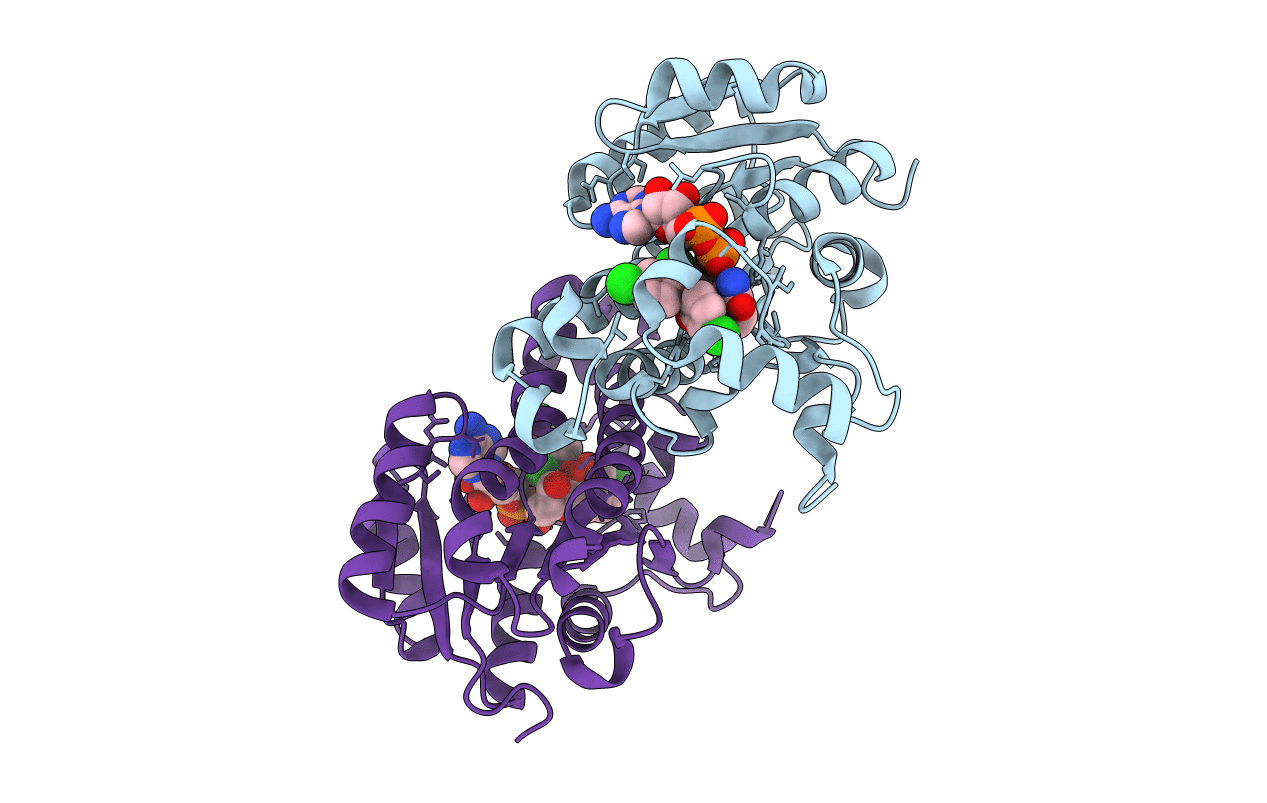
3NRC image
Deposition Date 2010-06-30
Release Date 2010-11-10
Last Version Date 2023-09-06
Entry Detail
PDB ID:
3NRC
Keywords:
Title:
Crystal Structure of the Francisella tularensis enoyl-acyl carrier protein reductase (FabI) in complex with NAD+ and triclosan
Biological Source:
Host Organism:
Method Details:
Experimental Method:
Resolution:
2.10 Å
R-Value Free:
0.24
R-Value Work:
0.18
R-Value Observed:
0.18
Space Group:
P 21 21 2
Macromolecular Entities
Polymer Type:polypeptide(L)
Molecule:Enoyl-[acyl-carrier-protein] reductase (NADH)
Gene (Uniprot):fabI
Chain IDs:A, B
Chain Length:280
Number of Molecules:2
Biological Source:Francisella tularensis subsp. tularensis
Primary Citation
Structure of the Francisella tularensis enoyl-acyl carrier protein reductase (FabI) in complex with NAD(+) and triclosan.
Acta Crystallogr.,Sect.F 66 1436 1440 (2010)
PMID: 21045289 DOI: 10.1107/S1744309110039862

Abstact

Enoyl-acyl carrier protein reductase (FabI) catalyzes the last rate-limiting step in the elongation cycle of the fatty-acid biosynthesis pathway and has been validated as a potential antimicrobial drug target in Francisella tularensis. The development of new antibiotic therapies is important both to combat potential drug-resistant bioweapons and to address the broader societal problem of increasing antibiotic resistance among many pathogenic bacteria. The crystal structure of FabI from F. tularensis (FtuFabI) in complex with the inhibitor triclosan and the cofactor NAD(+) has been solved to a resolution of 2.1 Å. Triclosan is known to effectively inhibit FabI from different organisms. Precise characterization of the mode of triclosan binding is required to develop highly specific inhibitors. Comparison of our structure with the previously determined FtuFabI structure (PDB code 2jjy) which is bound to only NAD(+) reveals the conformation of the substrate-binding loop, electron density for which was missing in the earlier structure, and demonstrates a shift in the conformation of the NAD(+) cofactor. This shift in the position of the phosphate groups allows more room in the active site for substrate or inhibitor to bind and be better accommodated. This information will be crucial for virtual screening studies to identify novel scaffolds for development into new active inhibitors.

Legend

Protein

Chemical

Disease

Primary Citation of related structures