3NPM image
Deposition Date 2010-06-28
Release Date 2011-05-11
Last Version Date 2023-09-06
Entry Detail
PDB ID:
3NPM
Keywords:
Title:
Crystal Structure of the C47A/A241C disulfide-linked C6 Aspartate Transcarbamoylase enzyme
Biological Source:
Source Organism:
Host Organism:
Method Details:
Experimental Method:
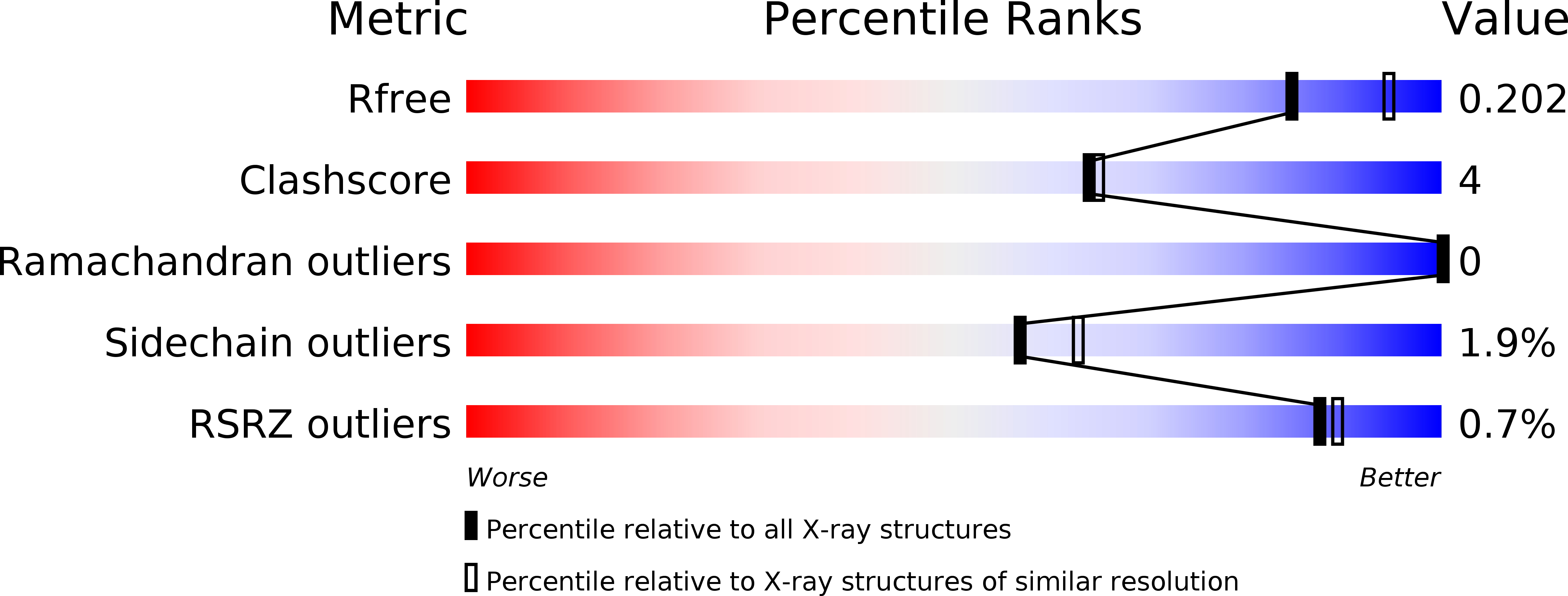
Resolution:
2.10 Å
R-Value Free:
0.20
R-Value Work:
0.17
R-Value Observed:
0.17
Space Group:
P 43 3 2
Macromolecular Entities
Structures with similar UniProt ID
Protein Blast
Polymer Type:polypeptide(L)
Molecule:Aspartate carbamoyltransferase catalytic chain
Gene (Uniprot):pyrB
Mutagens:A48C; C242A
Chain IDs:A
Chain Length:310
Number of Molecules:1
Biological Source:Escherichia coli
Ligand Molecules
Primary Citation
A cooperative Escherichia coli aspartate transcarbamoylase without regulatory subunits .
Biochemistry 49 7694 7703 (2010)
PMID: 20681545 DOI: 10.1021/bi1010333

Abstact

Here we report the isolation, kinetic characterization, and X-ray structure determination of a cooperative Escherichia coli aspartate transcarbamoylase (ATCase) without regulatory subunits. The native ATCase holoenzyme consists of six catalytic chains organized as two trimers bridged noncovalently by six regulatory chains organized as three dimers, c(6)r(6). Dissociation of the native holoenzyme produces catalytically active trimers, c(3), and nucleotide-binding regulatory dimers, r(2). By introducing specific disulfide bonds linking the catalytic chains from the upper trimer site specifically to their corresponding chains in the lower trimer prior to dissociation, a new catalytic unit, c(6), was isolated consisting of two catalytic trimers linked by disulfide bonds. Not only does the c(6) species display enhanced enzymatic activity compared to the wild-type enzyme, but the disulfide bonds also impart homotropic cooperativity, never observed in the wild-type c(3). The c(6) ATCase was crystallized in the presence of phosphate and its X-ray structure determined to 2.10 A resolution. The structure of c(6) ATCase liganded with phosphate exists in a nearly identical conformation as other R-state structures with similar values calculated for the vertical separation and planar angles. The disulfide bonds linking upper and lower catalytic trimers predispose the active site into a more active conformation by locking the 240s loop into the position characteristic of the high-affinity R state. Furthermore, the elimination of the structural constraints imposed by the regulatory subunits within the holoenzyme provides increased flexibility to the c(6) enzyme, enhancing its activity over the wild-type holoenzyme (c(6)r(6)) and c(3). The covalent linkage between upper and lower catalytic trimers restores homotropic cooperativity so that a binding event at one or so active sites stimulates binding at the other sites. Reduction of the disulfide bonds in the c(6) ATCase results in c(3) catalytic subunits that display kinetic parameters similar to those of wild-type c(3). This is the first report of an active c(6) catalytic unit that displays enhanced activity and homotropic cooperativity.

Legend

Protein

Chemical

Disease

Primary Citation of related structures