Deposition Date
1999-02-03
Release Date
2000-02-04
Last Version Date
2023-12-27
Entry Detail
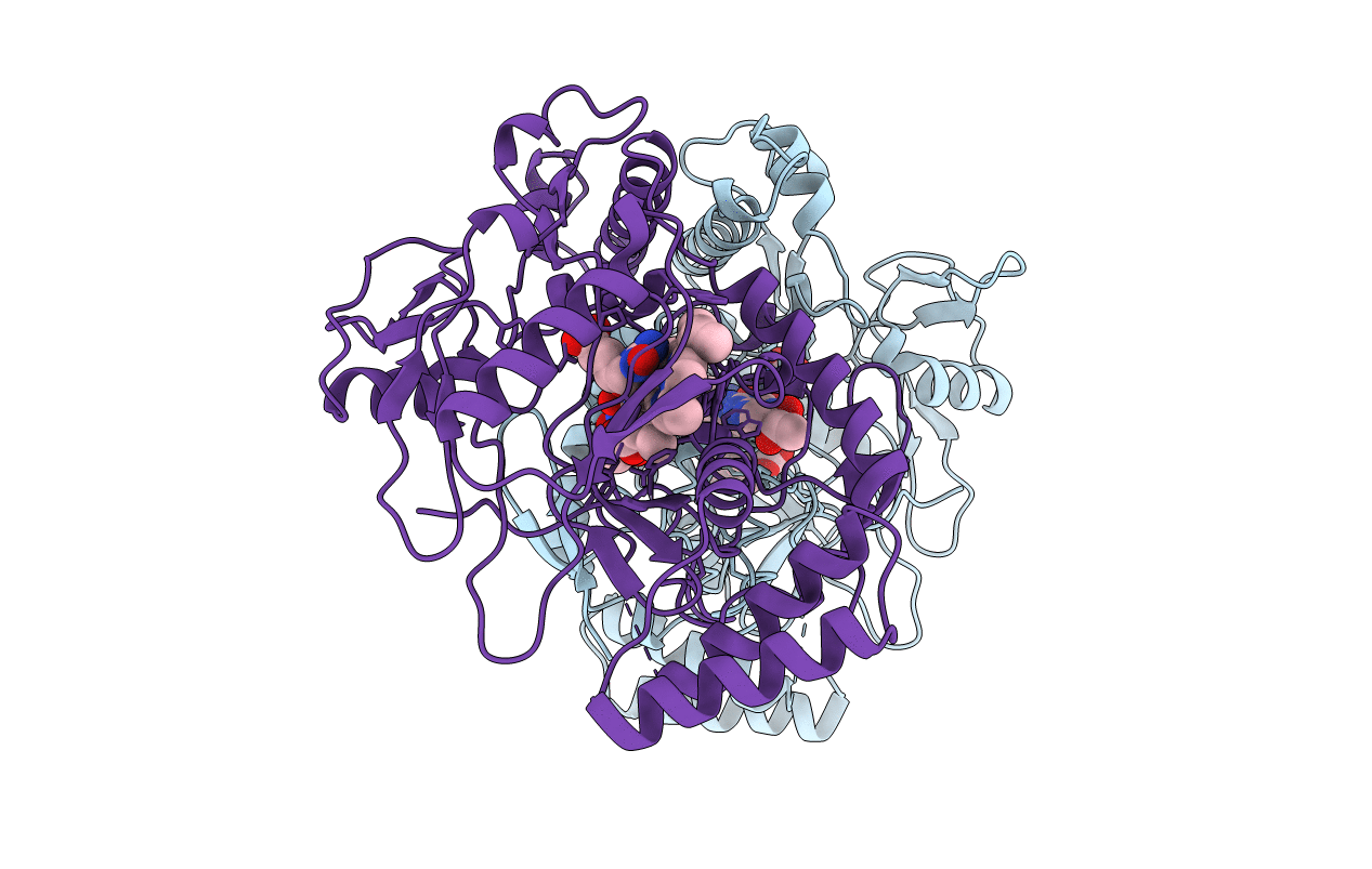
PDB ID:
3NOS
Keywords:
Title:
HUMAN ENDOTHELIAL NITRIC OXIDE SYNTHASE WITH ARGININE SUBSTRATE
Biological Source:
Source Organism(s):
Homo sapiens (Taxon ID: 9606)
Expression System(s):
Method Details:
Experimental Method:
Resolution:
2.40 Å
R-Value Free:
0.30
R-Value Work:
0.19
Space Group:
P 21 21 21