Deposition Date
2010-06-24
Release Date
2010-07-14
Last Version Date
2024-11-06
Entry Detail
PDB ID:
3NNQ
Keywords:
Title:
Crystal Structure of the N-terminal domain of Moloney murine leukemia virus integrase, Northeast Structural Genomics Consortium Target OR3
Biological Source:
Source Organism(s):
Moloney murine leukemia virus (Taxon ID: 11801)
Expression System(s):
Method Details:
Experimental Method:
Resolution:
2.69 Å
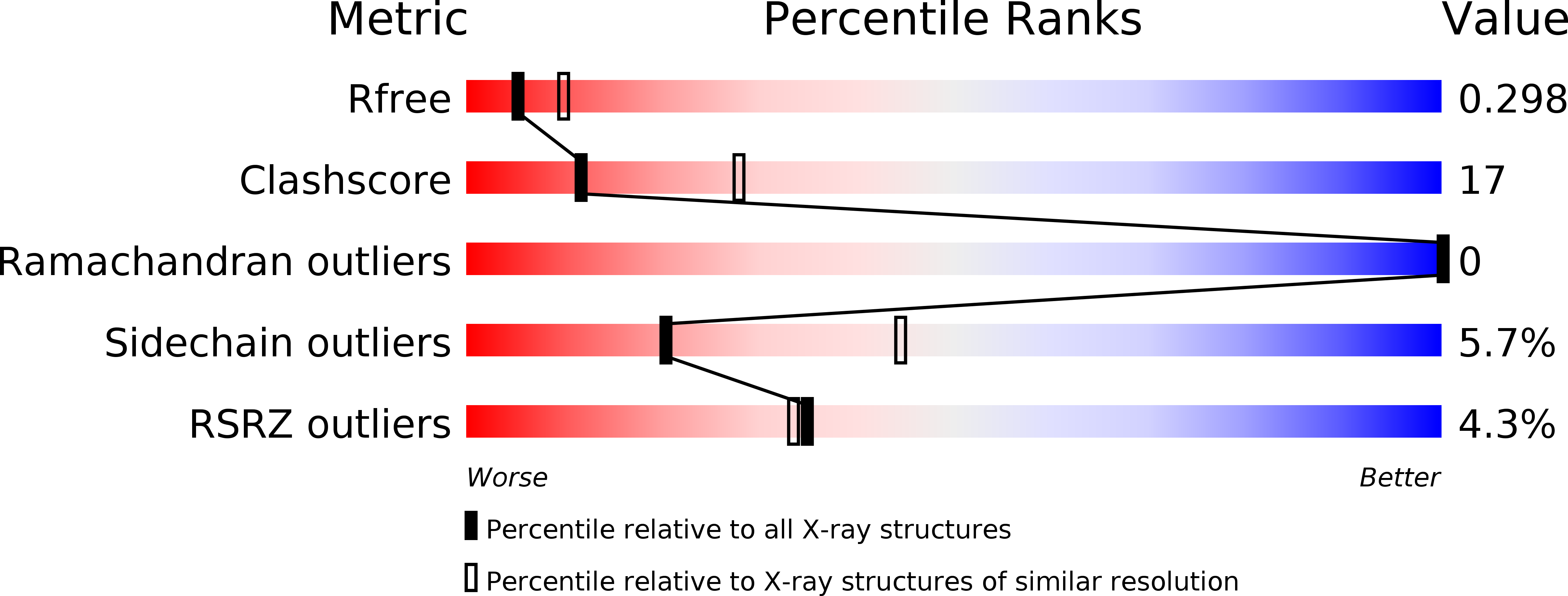
R-Value Free:
0.27
R-Value Work:
0.22
R-Value Observed:
0.23
Space Group:
H 3 2