3NMD image
Deposition Date 2010-06-22
Release Date 2010-09-08
Last Version Date 2024-10-30
Entry Detail
PDB ID:
3NMD
Keywords:
Title:
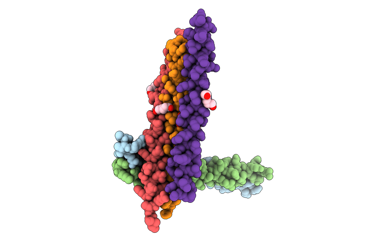
Crystal structure of the leucine zipper domain of cGMP dependent protein kinase I beta
Biological Source:
Source Organism(s):
Homo sapiens (Taxon ID: 9606)
Expression System(s):
Method Details:
Experimental Method:
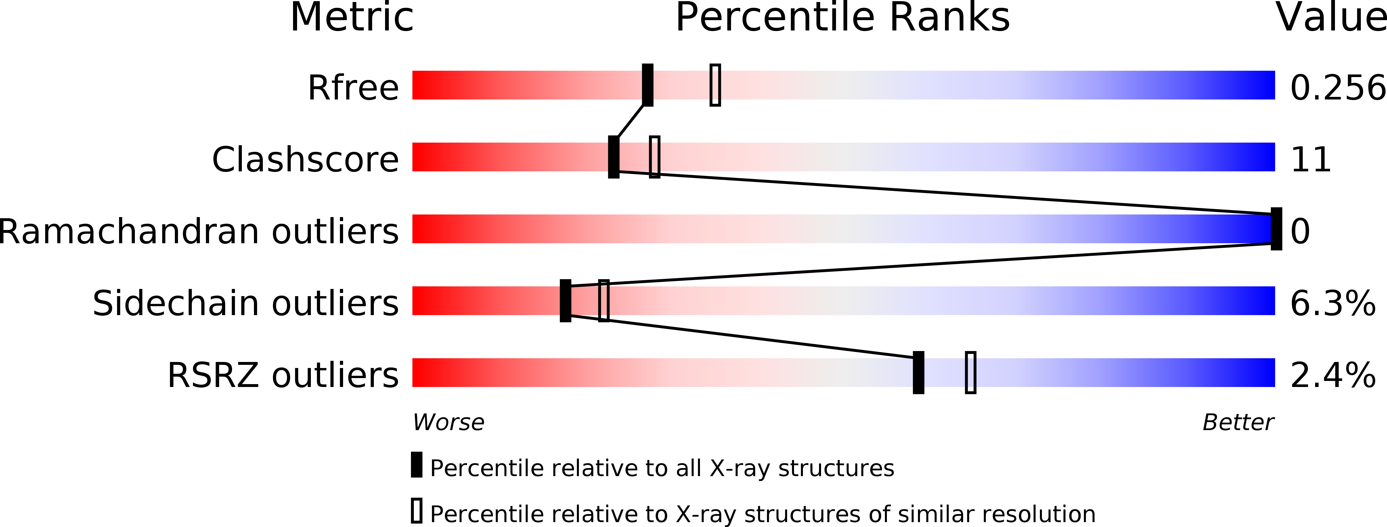
Resolution:
2.27 Å
R-Value Free:
0.24
R-Value Work:
0.19
R-Value Observed:
0.19
Space Group:
C 2 2 21
Macromolecular Entities
Structures with similar UniProt ID
Protein Blast
Polymer Type:polypeptide(L)
Molecule:cGMP Dependent PRotein Kinase
Mutagens:K41M
Chain IDs:A, B, C, D, E
Chain Length:72
Number of Molecules:5
Biological Source:Homo sapiens
Modified Residue
Compound ID Chain ID Parent Comp ID Details 2D Image
MSE A MET SELENOMETHIONINE
Primary Citation
A crystal structure of the cyclic GMP-dependent protein kinase I{beta} dimerization/docking domain reveals molecular details of isoform-specific anchoring.
J.Biol.Chem. 285 32684 32688 (2010)
PMID: 20826808 DOI: 10.1074/jbc.C110.161430

Abstact

Cyclic GMP-dependent protein kinase (PKG) is a key mediator of the nitric oxide/cGMP signaling pathway and plays a central role in regulating cardiovascular and neuronal functions. The N-terminal ∼50 amino acids of the kinase are required for homodimerization and association with isoform-specific PKG-anchoring proteins (GKAPs), which target the kinase to specific substrates. To understand the molecular details of PKG dimerization and gain insight into its association with GKAPs, we solved a crystal structure of the PKG Iβ dimerization/docking domain. Our structure provides molecular details of this unique leucine/isoleucine zipper, revealing specific hydrophobic and ionic interactions that mediate dimerization and demonstrating the topology of the GKAP interaction surface.

Legend

Protein

Chemical

Disease

Primary Citation of related structures
Feedback Form
Name
Email
Institute
Feedback