Deposition Date
2010-06-21
Release Date
2010-07-21
Last Version Date
2024-10-09
Entry Detail
PDB ID:
3NL9
Keywords:
Title:
Crystal structure of a putative NTP pyrophosphohydrolase (Exig_1061) from EXIGUOBACTERIUM SP. 255-15 at 1.78 A resolution
Biological Source:
Source Organism(s):
Exiguobacterium sibiricum 255-15 (Taxon ID: 262543)
Expression System(s):
Method Details:
Experimental Method:
Resolution:
1.78 Å
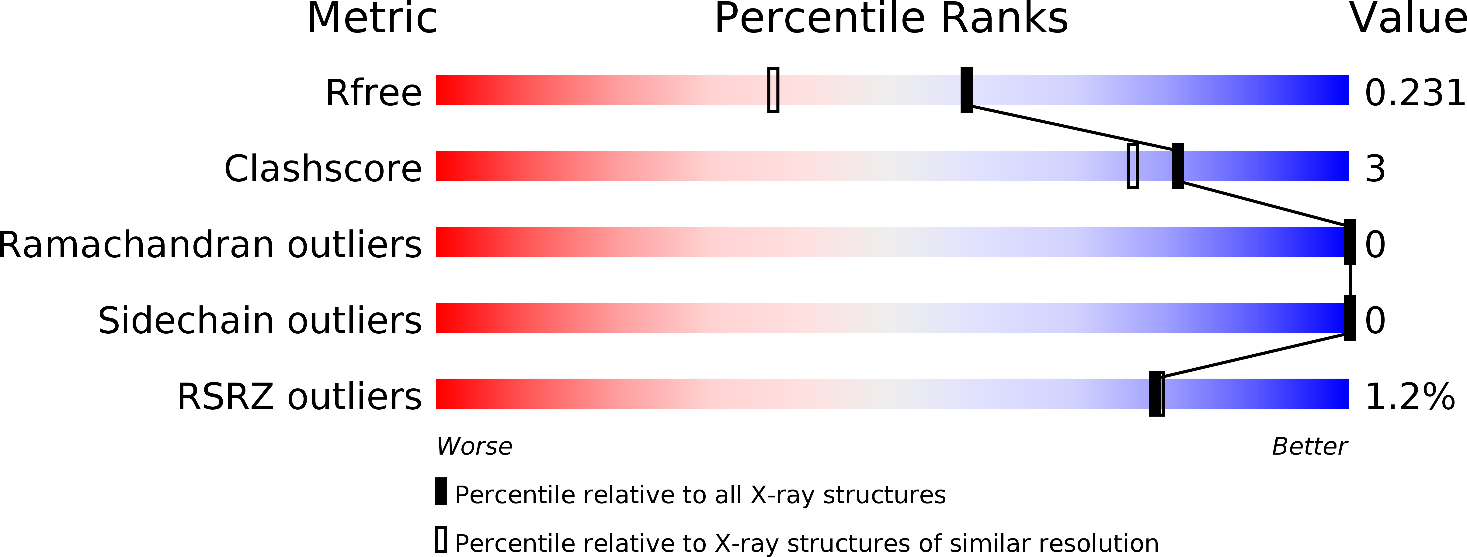
R-Value Free:
0.22
R-Value Work:
0.17
R-Value Observed:
0.17
Space Group:
C 1 2 1