Deposition Date
2010-06-16
Release Date
2011-06-01
Last Version Date
2023-11-01
Entry Detail
PDB ID:
3NIQ
Keywords:
Title:
Crystal structure of Pseudomonas aeruginosa guanidinopropionase
Biological Source:
Source Organism(s):
Pseudomonas aeruginosa (Taxon ID: 208964)
Expression System(s):
Method Details:
Experimental Method:
Resolution:
2.07 Å
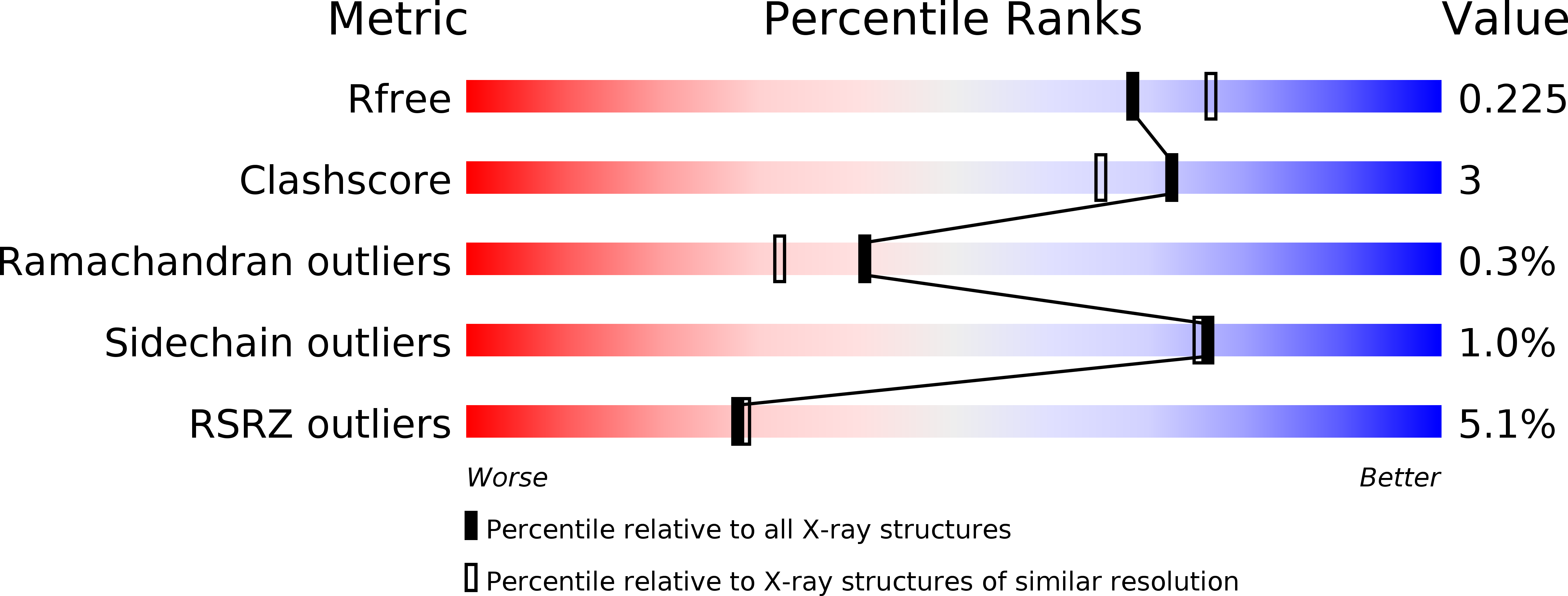
R-Value Free:
0.22
R-Value Work:
0.18
R-Value Observed:
0.18
Space Group:
P 21 3