3NH7 image
Entry Detail
PDB ID:
3NH7
Keywords:
Title:
Crystal structure of the neutralizing Fab fragment AbD1556 bound to the BMP type I receptor IA
Biological Source:
Source Organism:
Host Organism:
PDB Version:
Deposition Date:
2010-06-14
Release Date:
2010-10-27
Method Details:
Experimental Method:
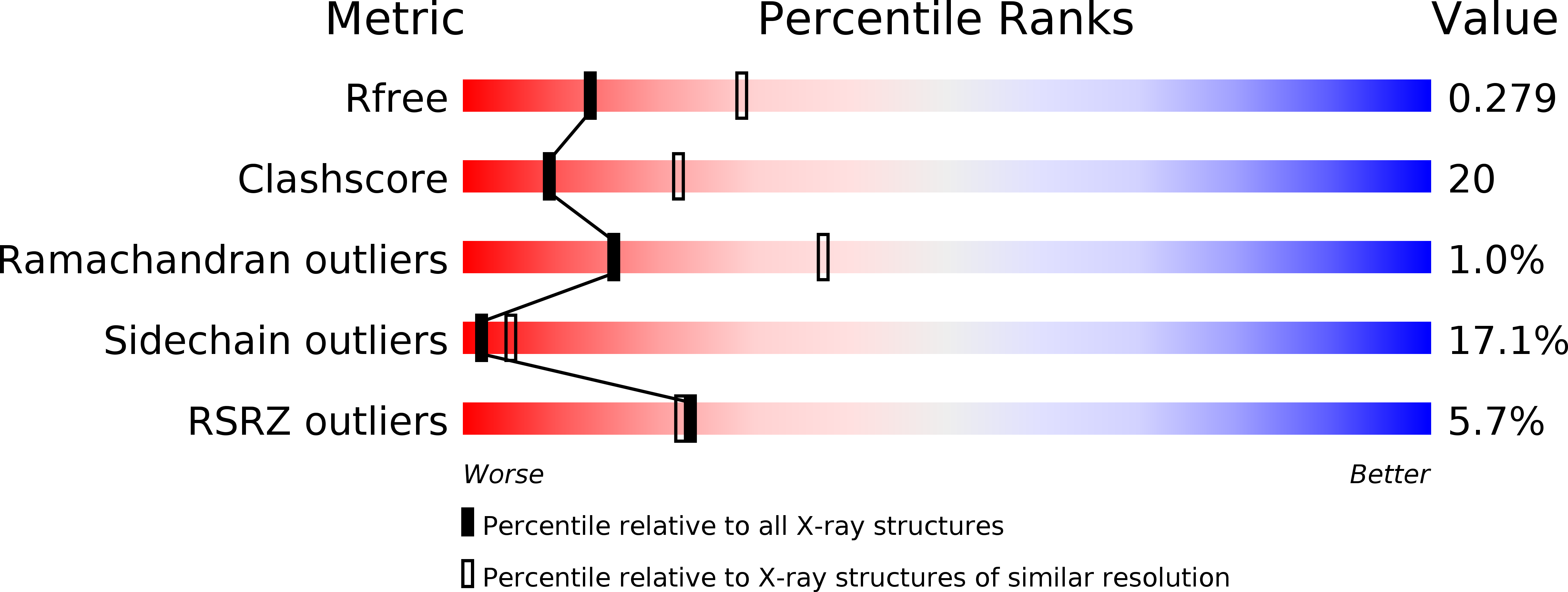
Resolution:
2.70 Å
R-Value Free:
0.28
R-Value Work:
0.23
R-Value Observed:
0.23
Space Group:
P 1 21 1
Macromolecular Entities
Polymer Type:polypeptide(L)
Description:Bone morphogenetic protein receptor type-1A
Chain IDs:C (auth: A), F (auth: B), I (auth: C), L (auth: D)
Chain Length:129
Number of Molecules:4
Biological Source:Homo sapiens
Polymer Type:polypeptide(L)
Description:Antibody fragment Fab AbD1556, heavy chain
Chain IDs:A (auth: H), D (auth: I), G (auth: J), J (auth: K)
Chain Length:234
Number of Molecules:4
Biological Source:Homo sapiens
Polymer Type:polypeptide(L)
Description:Antibody fragment Fab AbD1556, light chain
Chain IDs:B (auth: L), E (auth: M), H (auth: N), K (auth: O)
Chain Length:213
Number of Molecules:4
Biological Source:Homo sapiens
Primary Citation
A selection fit mechanism in BMP receptor IA as a possible source for BMP ligand-receptor promiscuity
Plos One 5 e13049 e13049 (2010)
PMID: 20927405 DOI: 10.1371/journal.pone.0013049

Abstact

BACKGROUND Members of the TGF-β superfamily are characterized by a highly promiscuous ligand-receptor interaction as is readily apparent from the numeral discrepancy of only seven type I and five type II receptors available for more than 40 ligands. Structural and functional studies have been used to address the question of how specific signals can be deduced from a limited number of receptor combinations and to unravel the molecular mechanisms underlying the protein-protein recognition that allow such limited specificity. PRINCIPAL FINDINGS In this study we have investigated how an antigen binding antibody fragment (Fab) raised against the extracellular domain of the BMP receptor type IA (BMPR-IA) recognizes the receptor's BMP-2 binding epitope and thereby neutralizes BMP-2 receptor activation. The crystal structure of the complex of the BMPR-IA ectodomain bound to the Fab AbD1556 revealed that the contact surface of BMPR-IA overlaps extensively with the contact surface for BMP-2 interaction. Although the structural epitopes of BMPR-IA to both binding partners coincides, the structures of BMPR-IA in the two complexes differ significantly. In contrast to the structural differences, alanine-scanning mutagenesis of BMPR-IA showed that the functional determinants for binding to the antibody and BMP-2 are almost identical. CONCLUSIONS Comparing the structures of BMPR-IA bound to BMP-2 or bound to the Fab AbD1556 with the structure of unbound BMPR-IA shows that binding of BMPR-IA to its interaction partners follows a selection fit mechanism, possibly indicating that the ligand promiscuity of BMPR-IA is inherently encoded by structural adaptability. The functional and structural analysis of the BMPR-IA binding antibody AbD1556 mimicking the BMP-2 binding epitope may thus pave the way for the design of low-molecular weight synthetic receptor binders/inhibitors.

Legend

Protein

Chemical

Disease

Primary Citation of related structures