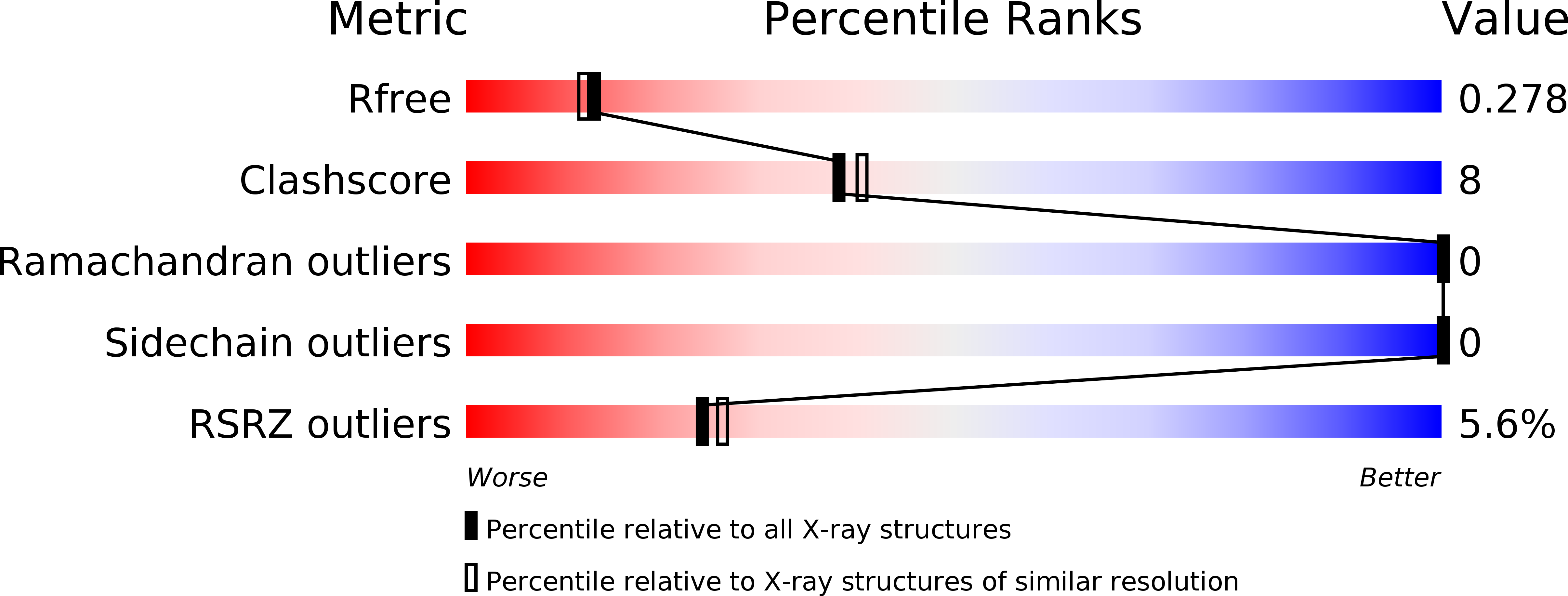
3NGK image
Deposition Date 2010-06-11
Release Date 2010-10-06
Last Version Date 2023-09-06
Entry Detail
PDB ID:
3NGK
Title:
PduA from Salmonella enterica Typhimurium
Biological Source:
Method Details:
Experimental Method:
Resolution:
2.26 Å
R-Value Free:
0.28
R-Value Work:
0.25
R-Value Observed:
0.25
Space Group:
P 6 2 2
Macromolecular Entities
Polymer Type:polypeptide(L)
Molecule:Propanediol utilization protein pduA
Gene (Uniprot):pduA
Chain IDs:A
Chain Length:102
Number of Molecules:1
Biological Source:Salmonella enterica subsp. enterica serovar Typhimurium
Primary Citation
Structural Insight into the Mechanisms of Transport across the Salmonella enterica Pdu Microcompartment Shell.
J.Biol.Chem. 285 37838 37846 (2010)
PMID: 20870711 DOI: 10.1074/jbc.M110.160580

Abstact

Bacterial microcompartments are a functionally diverse group of proteinaceous organelles that confine specific reaction pathways in the cell within a thin protein-based shell. The propanediol utilizing (Pdu) microcompartment contains the reactions for metabolizing 1,2-propanediol in certain enteric bacteria, including Salmonella. The Pdu shell is assembled from a few thousand protein subunits of several different types. Here we report the crystal structures of two key shell proteins, PduA and PduT. The crystal structures offer insights into the mechanisms of Pdu microcompartment assembly and molecular transport across the shell. PduA forms a symmetric homohexamer whose central pore appears tailored for facilitating transport of the 1,2-propanediol substrate. PduT is a novel, tandem domain shell protein that assembles as a pseudohexameric homotrimer. Its structure reveals an unexpected site for binding an [Fe-S] cluster at the center of the PduT pore. The location of a metal redox cofactor in the pore of a shell protein suggests a novel mechanism for either transferring redox equivalents across the shell or for regenerating luminal [Fe-S] clusters.

Legend

Protein

Chemical

Disease

Primary Citation of related structures