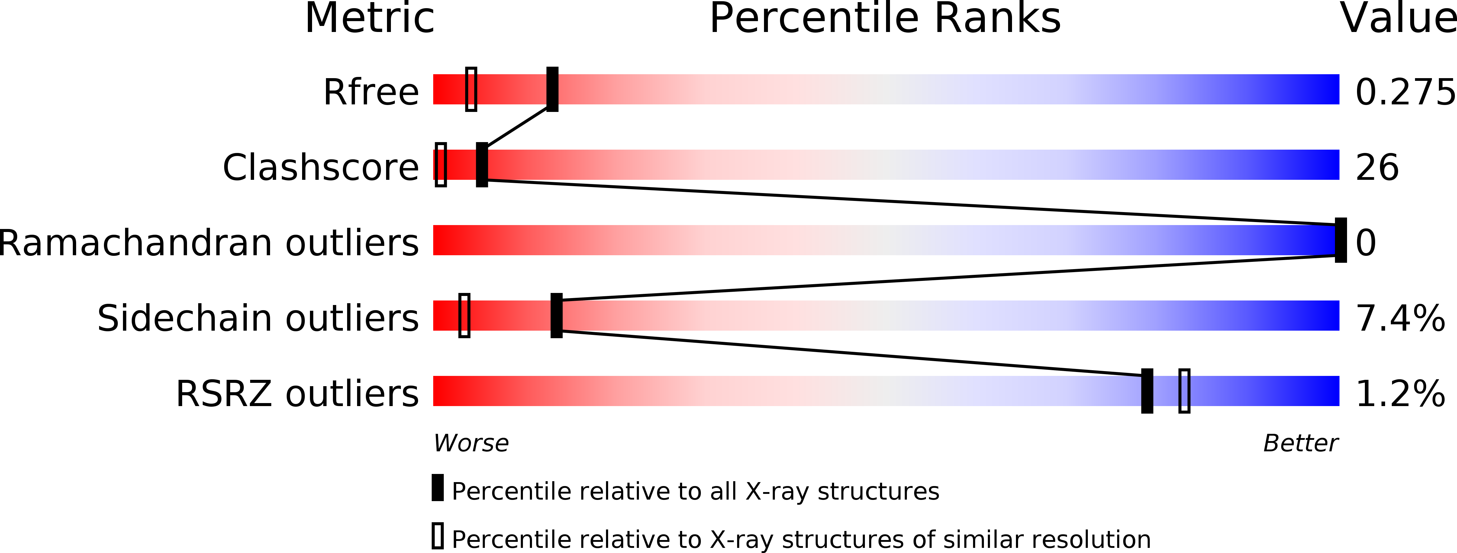
3NFY image
Deposition Date 2010-06-10
Release Date 2010-11-10
Last Version Date 2023-09-06
Entry Detail
PDB ID:
3NFY
Keywords:
Title:
The Structure of Human Bisphosphoglycerate Mutase to 1.94A
Biological Source:
Source Organism:
Homo sapiens (Taxon ID: 9606)
Host Organism:
Method Details:
Experimental Method:
Resolution:
1.94 Å
R-Value Free:
0.27
R-Value Work:
0.17
R-Value Observed:
0.18
Space Group:
P 1 21 1
Macromolecular Entities
Structures with similar UniProt ID
Protein Blast
Polymer Type:polypeptide(L)
Molecule:Bisphosphoglycerate mutase
Gene (Uniprot):BPGM
Chain IDs:A, B
Chain Length:267
Number of Molecules:2
Biological Source:Homo sapiens
Primary Citation
Unliganded structure of human bisphosphoglycerate mutase reveals side-chain movements induced by ligand binding.
Acta Crystallogr.,Sect.F 66 1415 1420 (2010)
PMID: 21045285 DOI: 10.1107/S1744309110035475

Abstact

Erythrocyte-specific bisphosphoglycerate mutase is a trifunctional enzyme which modulates the levels of 2,3-bisphosphoglycerate (2,3-BPG) in red blood cells by virtue of its synthase and phosphatase activities. Low levels of erythrocyte 2,3-BPG increase the affinity of haemoglobin for oxygen, thus limiting the release of oxygen into tissues. 2,3-BPG levels in stored blood decline rapidly owing to the phosphatase activity of bisphosphoglycerate mutase, which is enhanced by a fall in pH. Here, the 1.94 Å resolution X-ray structure of bisphosphoglycerate mutase is presented, focusing on the dynamic nature of key ligand-binding residues and their interaction with the inhibitor citrate. Residues at the binding pocket are complete. In addition, the movement of key residues in the presence and absence of ligand is described and alternative conformations are explored. The conformation in which the ligand citrate would bind at the substrate-binding pocket is proposed, with discussion and representations of its orientation. The characterization of bisphosphoglycerate mutase-citrate interactions will provide a framework for the design of specific inhibitors of the phosphatase activity of this enzyme, which may limit the decline of 2,3-BPG in stored blood.

Legend

Protein

Chemical

Disease

Primary Citation of related structures