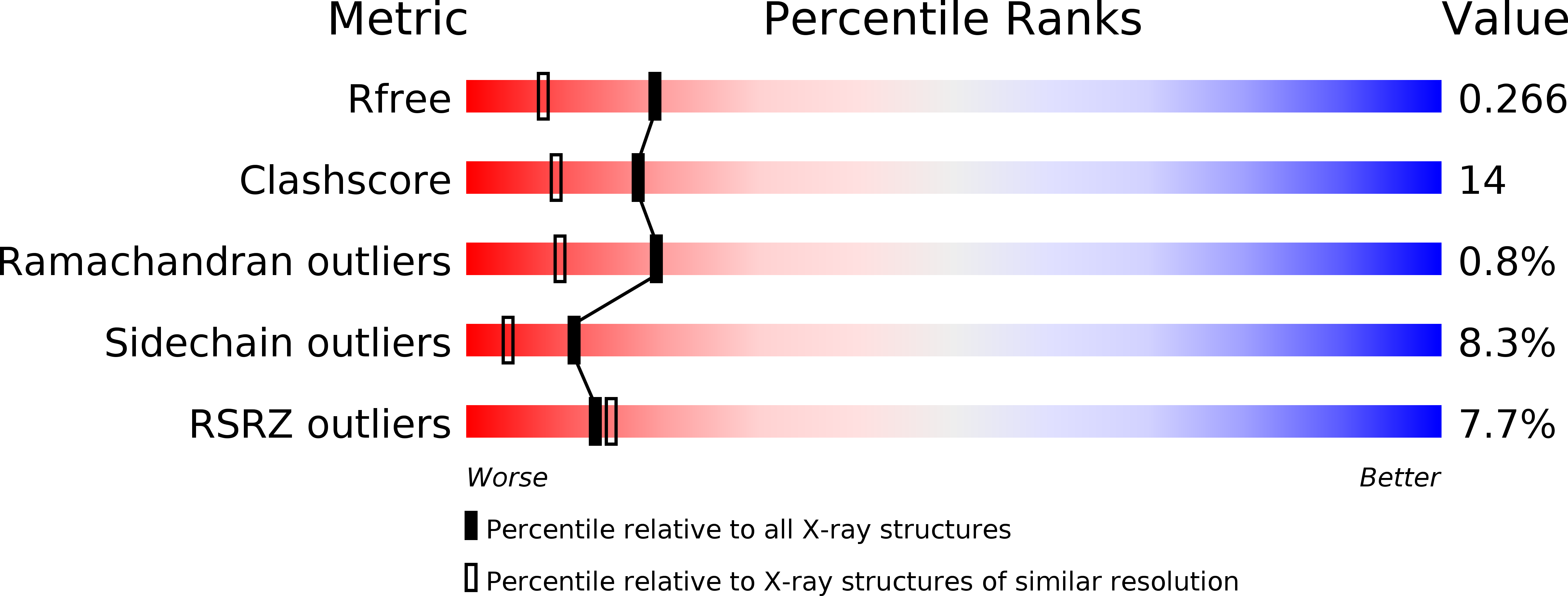
3NFD image
Deposition Date 2010-06-10
Release Date 2011-01-05
Last Version Date 2024-11-27
Entry Detail
PDB ID:
3NFD
Keywords:
Title:
Chronobacterium ammoniagenes ACPS-CoA complex
Biological Source:
Source Organism:
Host Organism:
Method Details:
Experimental Method:
Resolution:
1.89 Å
R-Value Free:
0.25
R-Value Work:
0.21
R-Value Observed:
0.21
Space Group:
P 1
Macromolecular Entities
Polymer Type:polypeptide(L)
Molecule:Phosphopantetheine protein transferase, Ppt1p
Gene (Uniprot):PPT1
Chain IDs:A, B, C, D, E, F
Chain Length:153
Number of Molecules:6
Biological Source:Corynebacterium ammoniagenes
Modified Residue
Compound ID Chain ID Parent Comp ID Details 2D Image
MSE A MET SELENOMETHIONINE
Ligand Molecules
Primary Citation
Mycobacterium tuberculosis acyl carrier protein synthase adopts two different pH-dependent structural conformations.
Acta Crystallogr.,Sect.D 67 657 669 (2011)
PMID: 21697604 DOI: 10.1107/S0907444911020221

Abstact

The crystal structures of acyl carrier protein synthase (AcpS) from Mycobacterium tuberculosis (Mtb) and Corynebacterium ammoniagenes determined at pH 5.3 and pH 6.5, respectively, are reported. Comparison of the Mtb apo-AcpS structure with the recently reported structure of the Mtb AcpS-ADP complex revealed that AcpS adopts two different conformations: the orthorhombic and trigonal space-group structures show structural differences in the α2 helix and in the conformation of the α3-α4 connecting loop, which is in a closed conformation. The apo-AcpS structure shows electron density for the entire model and was obtained at lower pH values (4.4-6.0). In contrast, at a higher pH value (6.5) AcpS undergoes significant conformational changes, resulting in disordered regions that show no electron density in the AcpS model. The solved structures also reveal that C. ammoniagenes AcpS undergoes structural rearrangement in two regions, similar to the recently reported Mtb AcpS-ADP complex structure. In vitro reconstitution experiments show that AcpS has a higher post-translational modification activity between pH 4.4 and 6.0 than at pH values above 6.5, where the activity drops owing to the change in conformation. The results show that apo-AcpS and AcpS-ADP adopt different conformations depending upon the pH conditions of the crystallization solution.

Legend

Protein

Chemical

Disease

Primary Citation of related structures