Deposition Date
2010-06-09
Release Date
2011-04-13
Last Version Date
2024-11-20
Entry Detail
PDB ID:
3NEV
Keywords:
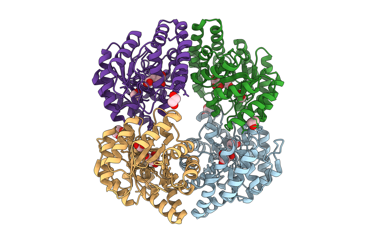
Title:
Crystal structure of YagE, a prophage protein from E. coli K12 in complex with KDGal
Biological Source:
Source Organism(s):
Escherichia coli (Taxon ID: 83333)
Expression System(s):
Method Details:
Experimental Method:
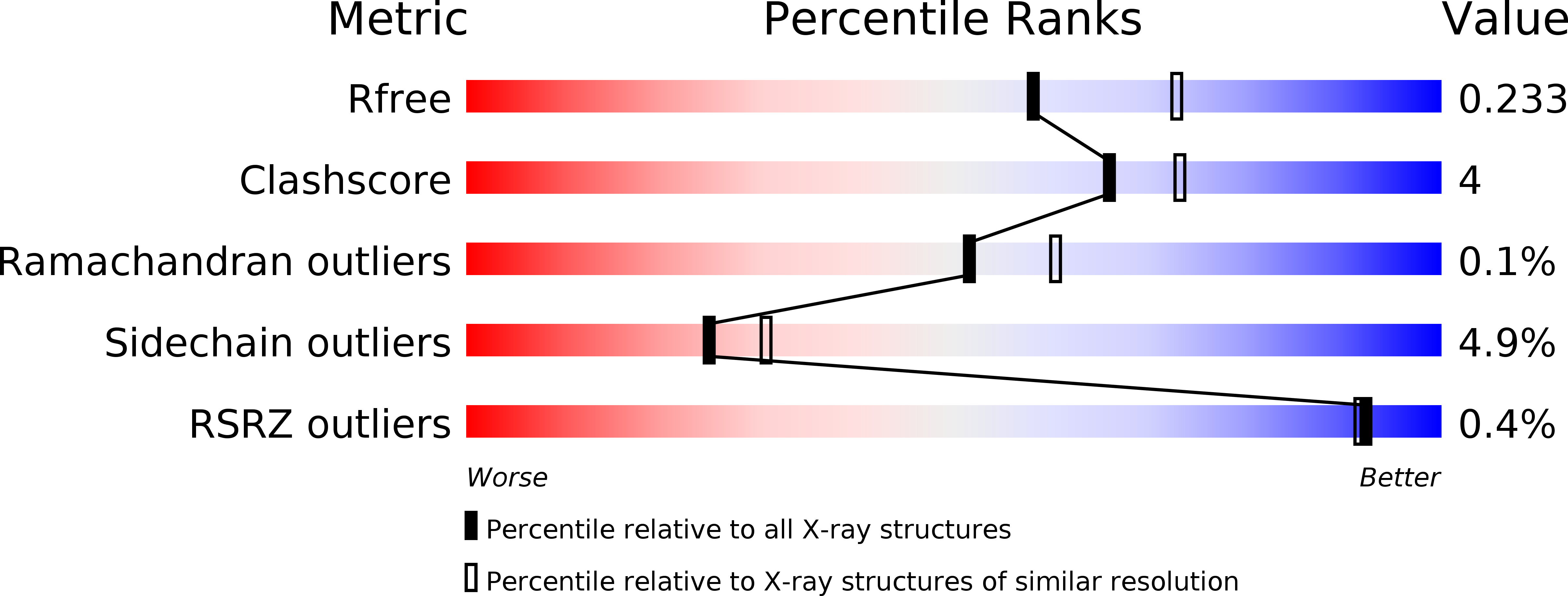
Resolution:
2.19 Å
R-Value Free:
0.23
R-Value Work:
0.19
R-Value Observed:
0.19
Space Group:
P 21 21 2