Deposition Date
2010-06-08
Release Date
2011-03-02
Last Version Date
2024-02-21
Entry Detail
PDB ID:
3NE5
Keywords:
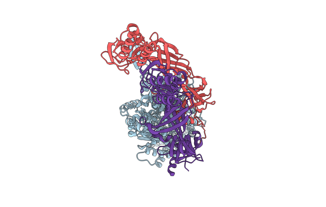
Title:
Crystal structure of the CusBA heavy-metal efflux complex from Escherichia coli
Biological Source:
Source Organism(s):
Escherichia coli (Taxon ID: 83333)
Expression System(s):
Method Details:
Experimental Method:
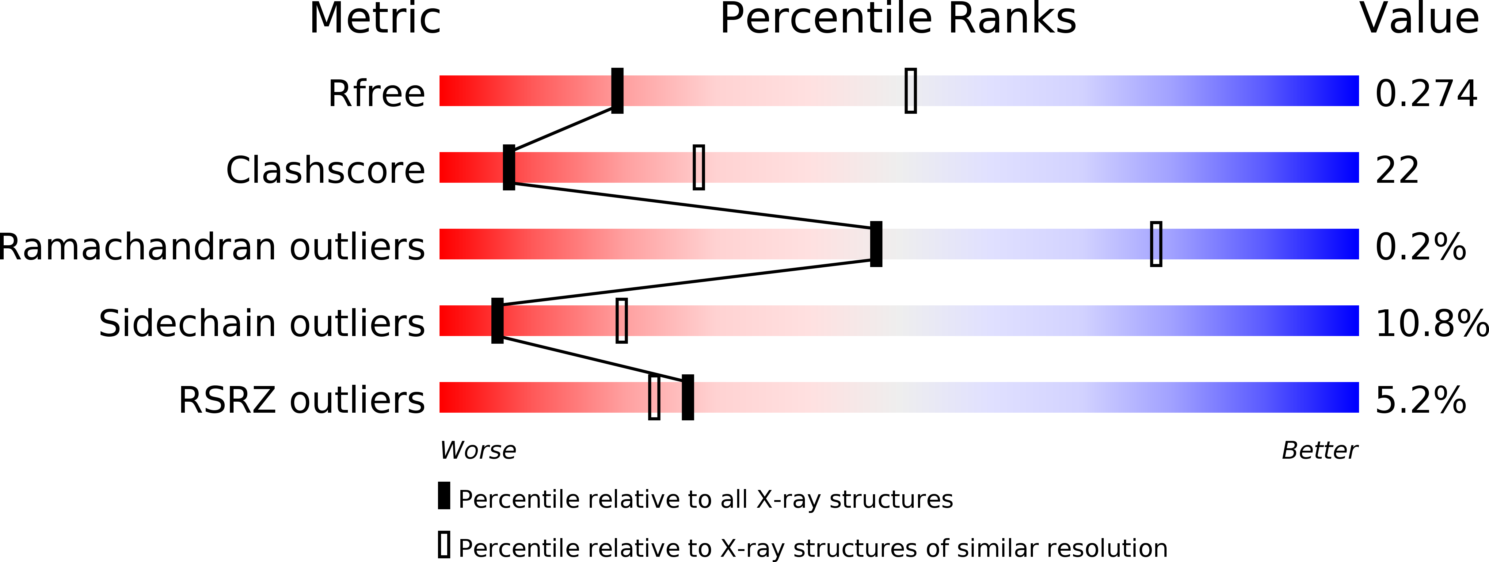
Resolution:
2.90 Å
R-Value Free:
0.26
R-Value Work:
0.22
R-Value Observed:
0.23
Space Group:
H 3 2