3NBX image
Deposition Date 2010-06-04
Release Date 2011-01-12
Last Version Date 2024-02-21
Entry Detail
PDB ID:
3NBX
Keywords:
Title:
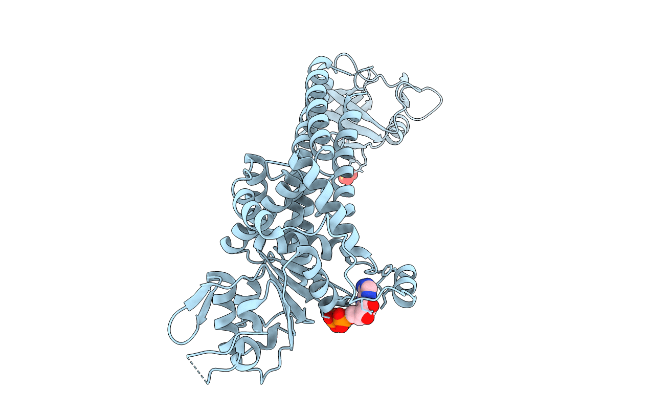
Crystal structure of E. coli RavA (Regulatory ATPase variant A) in complex with ADP
Biological Source:
Source Organism:
Host Organism:
Method Details:
Experimental Method:
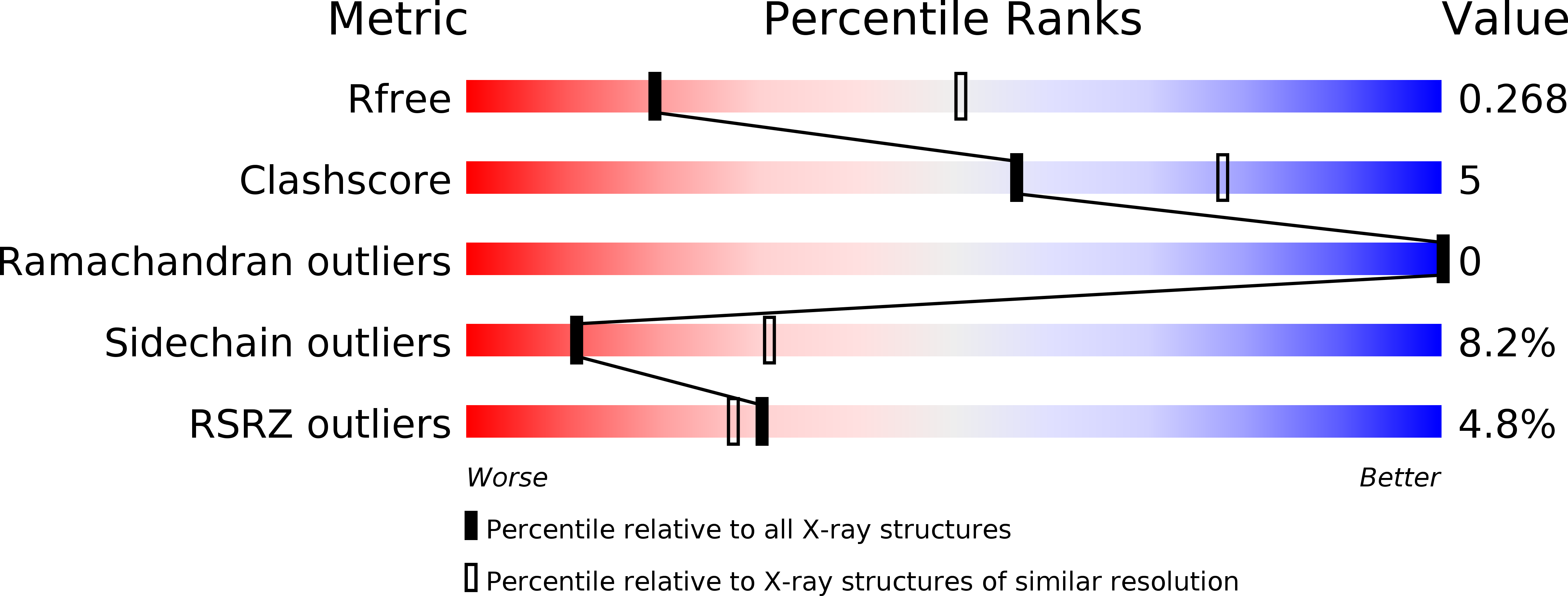
Resolution:
2.91 Å
R-Value Free:
0.26
R-Value Work:
0.22
R-Value Observed:
0.22
Space Group:
P 65
Macromolecular Entities
Structures with similar UniProt ID
Protein Blast
Polymer Type:polypeptide(L)
Molecule:ATPase ravA
Gene (Uniprot):ravA
Chain IDs:A (auth: X)
Chain Length:500
Number of Molecules:1
Biological Source:Escherichia coli
Primary Citation
Structure of RavA MoxR AAA+ protein reveals the design principles of a molecular cage modulating the inducible lysine decarboxylase activity
Proc.Natl.Acad.Sci.USA 107 22499 22504 (2010)
PMID: 21148420 DOI: 10.1073/pnas.1009092107

Abstact

The MoxR family of AAA+ ATPases is widespread throughout bacteria and archaea but remains poorly characterized. We recently found that the Escherichia coli MoxR protein, RavA (Regulatory ATPase variant A), tightly interacts with the inducible lysine decarboxylase, LdcI/CadA, to form a unique cage-like structure. Here, we present the X-ray structure of RavA and show that the αβα and all-α subdomains in the RavA AAA+ module are arranged as in magnesium chelatases rather than as in classical AAA+ proteins. RavA structure also contains a discontinuous triple-helical domain as well as a β-barrel-like domain forming a unique fold, which we termed the LARA domain. The LARA domain was found to mediate the interaction between RavA and LdcI. The RavA structure provides insights into how five RavA hexamers interact with two LdcI decamers to form the RavA-LdcI cage-like structure.

Legend

Protein

Chemical

Disease

Primary Citation of related structures
Feedback Form
Name
Email
Institute
Feedback