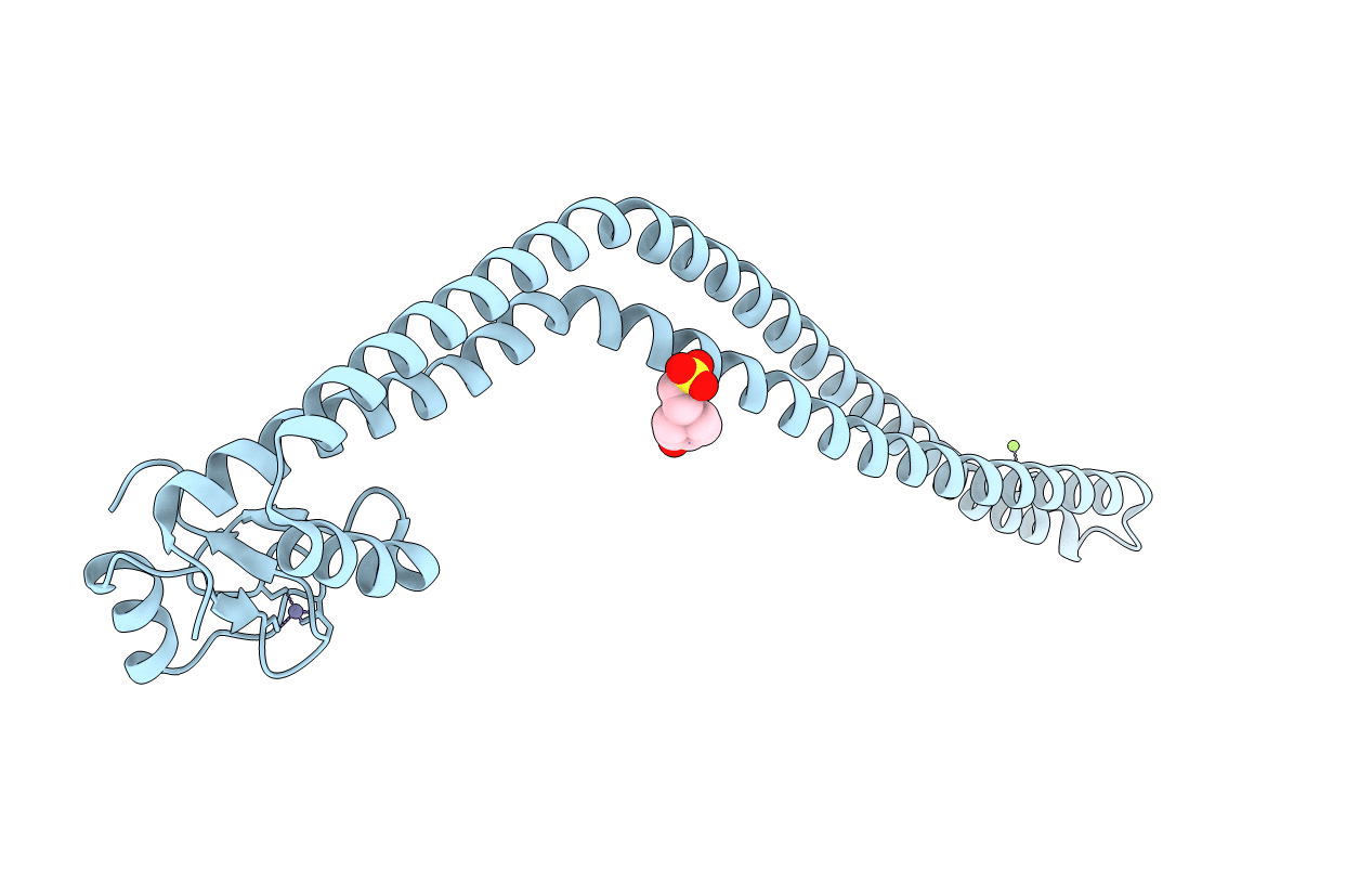
3NA7 image
Entry Detail
PDB ID:
3NA7
Title:
2.2 Angstrom Structure of the HP0958 Protein from Helicobacter pylori CCUG 17874
Biological Source:
Source Organism:
Host Organism:
PDB Version:
Deposition Date:
2010-06-01
Release Date:
2010-09-22
Method Details:
Experimental Method:
Resolution:
2.20 Å
R-Value Free:
0.29
R-Value Work:
0.24
R-Value Observed:
0.25
Space Group:
C 1 2 1
Macromolecular Entities
Polymer Type:polypeptide(L)
Description:HP0958
Chain IDs:A
Chain Length:256
Number of Molecules:1
Biological Source:Helicobacter pylori
Primary Citation
The 2.2-A Structure of the HP0958 Protein from Helicobacter pylori Reveals a Kinked Anti-Parallel Coiled-Coil Hairpin Domain and a Highly Conserved Zn-Ribbon Domain
J.Mol.Biol. 403 405 419 (2010)
PMID: 20826163 DOI: 10.1016/j.jmb.2010.08.051

Abstact

We have determined the 2.2-Å structure of the HP0958 protein from the human gastric pathogen Helicobacter pylori. HP0958 is essential for flagellum formation and motility. It functions as a chaperone for RpoN (σ(54)) and also controls the stability and translation of mRNA for the major flagellin subunit FlaA. The protein is composed of a highly elongated and kinked coiled-coil hairpin domain (residues 1-170), followed by a C(4) Zn-ribbon domain (residues 174-238). The Zn-ribbon domain is rich in aromatic and positively charged amino acid residues. Electrophoretic mobility shift assays identified residues in a positively charged region of the Zn-ribbon domain of HP0958 whose mutation alters the mobility of an HP0958-flaA mRNA complex. Mutation of surface residues in the coiled-coil domain did not result in an observable change in the mobility of the HP0958-flaA transcript complex. The data thus suggest the arrangement of HP0958 into distinct structural and functional domains.

Legend

Protein

Chemical

Disease

Primary Citation of related structures