3NA5 image
Deposition Date 2010-06-01
Release Date 2011-02-16
Last Version Date 2024-11-27
Entry Detail
PDB ID:
3NA5
Keywords:
Title:
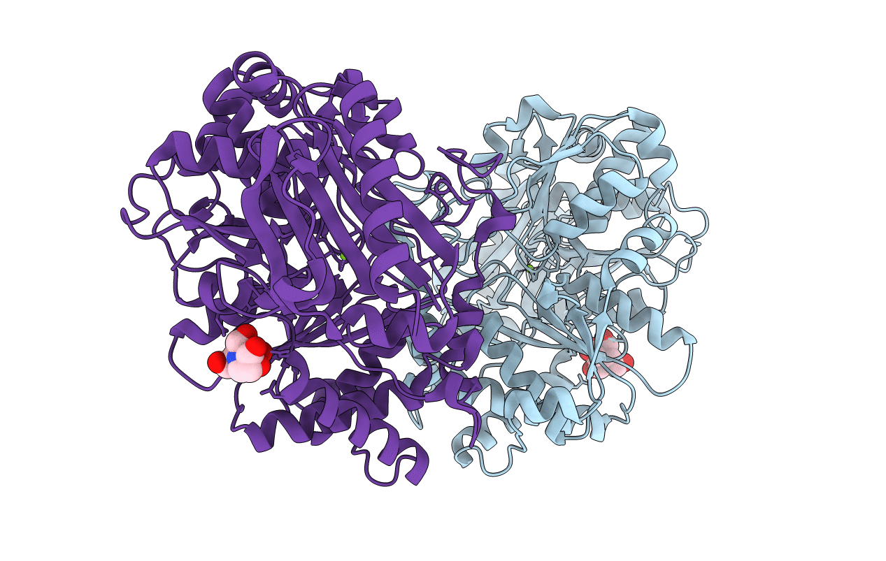
Crystal structure of a bacterial phosphoglucomutase, an enzyme important in the virulence of several human pathogens.
Biological Source:
Method Details:
Experimental Method:
Resolution:
1.70 Å
R-Value Free:
0.21
R-Value Work:
0.18
R-Value Observed:
0.18
Space Group:
P 21 21 21
Macromolecular Entities
Structures with similar UniProt ID
Protein Blast
Polymer Type:polypeptide(L)
Molecule:Phosphoglucomutase
Gene (Uniprot):pgm
Chain IDs:A, B
Chain Length:570
Number of Molecules:2
Biological Source:Salmonella enterica subsp. enterica serovar Typhimurium
Modified Residue
Compound ID Chain ID Parent Comp ID Details 2D Image
CSS A CYS S-MERCAPTOCYSTEINE
Primary Citation
Crystal structure of a bacterial phosphoglucomutase, an enzyme involved in the virulence of multiple human pathogens.
Proteins 79 1215 1229 (2011)
PMID: 21246636 DOI: 10.1002/prot.22957

Abstact

The crystal structure of the enzyme phosphoglucomutase from Salmonella typhimurium (StPGM) is reported at 1.7 A resolution. This is the first high-resolution structural characterization of a bacterial protein from this large enzyme family, which has a central role in metabolism and is also important to bacterial virulence and infectivity. A comparison of the active site of StPGM with that of other phosphoglucomutases reveals conserved residues that are likely involved in catalysis and ligand binding for the entire enzyme family. An alternate crystal form of StPGM and normal mode analysis give insights into conformational changes of the C-terminal domain that occur upon ligand binding. A novel observation from the StPGM structure is an apparent dimer in the asymmetric unit of the crystal, mediated largely through contacts in an N-terminal helix. Analytical ultracentrifugation and small-angle X-ray scattering confirm that StPGM forms a dimer in solution. Multiple sequence alignments and phylogenetic studies show that a distinct subset of bacterial PGMs share the signature dimerization helix, while other bacterial and eukaryotic PGMs are likely monomers. These structural, biochemical, and bioinformatic studies of StPGM provide insights into the large α-D-phosphohexomutase enzyme superfamily to which it belongs, and are also relevant to the design of inhibitors specific to the bacterial PGMs.

Legend

Protein

Chemical

Disease

Primary Citation of related structures