3NA0 image
Deposition Date 2010-05-31
Release Date 2010-09-08
Last Version Date 2024-02-21
Entry Detail
PDB ID:
3NA0
Title:
Crystal structure of human CYP11A1 in complex with 20,22-dihydroxycholesterol
Biological Source:
Source Organism:
Homo sapiens (Taxon ID: 9606)
Host Organism:
Method Details:
Experimental Method:
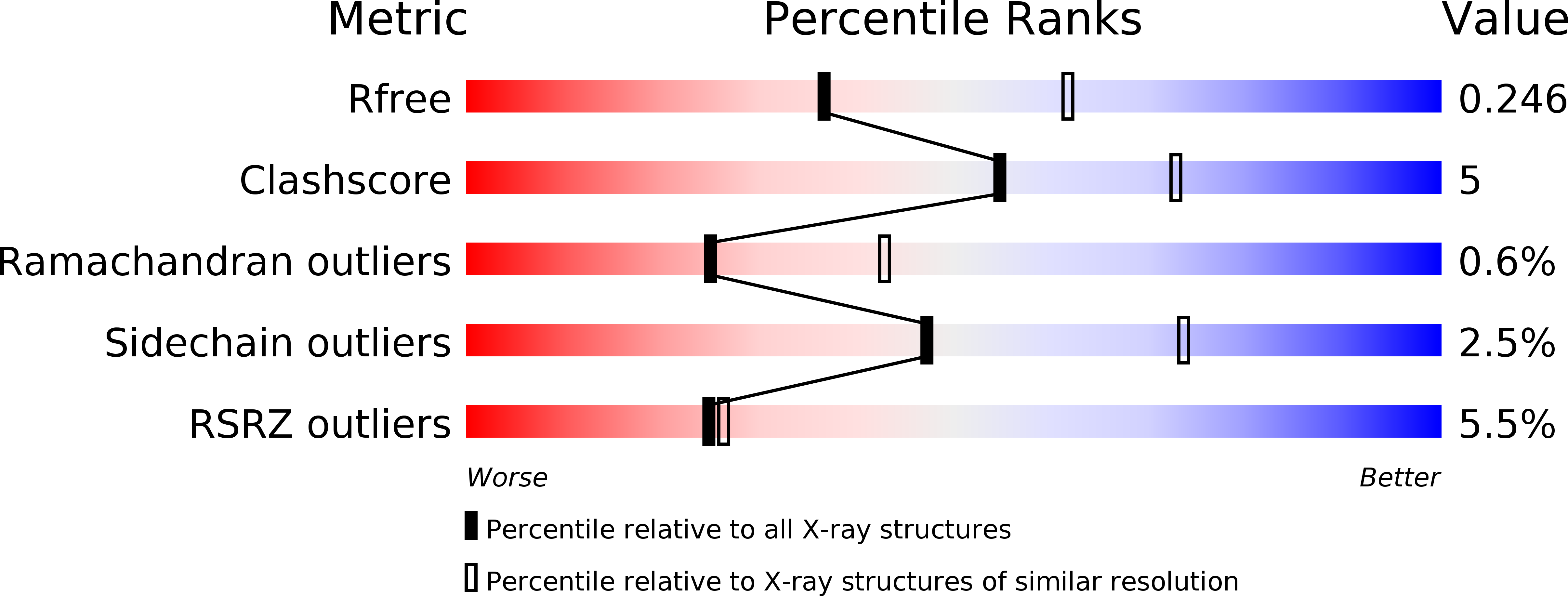
Resolution:
2.50 Å
R-Value Free:
0.24
R-Value Work:
0.20
R-Value Observed:
0.20
Space Group:
P 1 21 1
Macromolecular Entities
Polymer Type:polypeptide(L)
Molecule:Cholesterol side-chain cleavage enzyme, mitochondrial
Gene (Uniprot):CYP11A1
Chain IDs:A, B
Chain Length:471
Number of Molecules:2
Biological Source:Homo sapiens
Polymer Type:polypeptide(L)
Molecule:Adrenodoxin, mitochondrial
Gene (Uniprot):FDX1
Chain IDs:C, D
Chain Length:68
Number of Molecules:2
Biological Source:Homo sapiens
Primary Citation
Structural basis for pregnenolone biosynthesis by the mitochondrial monooxygenase system.
Proc.Natl.Acad.Sci.USA 108 10139 10143 (2011)
PMID: 21636783 DOI: 10.1073/pnas.1019441108

Abstact

In humans, the precursor to all steroid hormones, pregnenolone, is synthesized from cholesterol by an enzyme complex comprising adrenodoxin reductase (AdR), adrenodoxin (Adx), and a cytochrome P450 (P450scc or CYP11A1). This complex not only plays a key role in steroidogenesis, but also has long been a model to study electron transfer, multistep catalysis, and C-C bond cleavage performed by monooxygenases. Detailed mechanistic understanding of these processes has been hindered by a lack of structural information. Here we present the crystal structure of the complex of human Adx and CYP11A1--the first of a complex between a eukaryotic CYP and its redox partner. The structures with substrate and a series of reaction intermediates allow us to define the mechanism underlying sequential hydroxylations of the cholesterol and suggest the mechanism of C-C bond cleavage. In the complex the [2Fe-2S] cluster of Adx is positioned 17.4 Å away from the heme iron of CYP11A1. This structure suggests that after an initial protein-protein association driven by electrostatic forces, the complex adopts an optimized geometry between the redox centers. Conservation of the interaction interface suggests that this mechanism is common for all mitochondrial P450s.

Legend

Protein

Chemical

Disease

Primary Citation of related structures