Deposition Date
2010-05-28
Release Date
2010-08-11
Last Version Date
2023-09-06
Entry Detail
PDB ID:
3N9D
Keywords:
Title:
Monoclinic Structure of P. aeruginosa LigD phosphoesterase domain
Biological Source:
Source Organism(s):
Pseudomonas aeruginosa (Taxon ID: 287)
Expression System(s):
Method Details:
Experimental Method:
Resolution:
2.30 Å
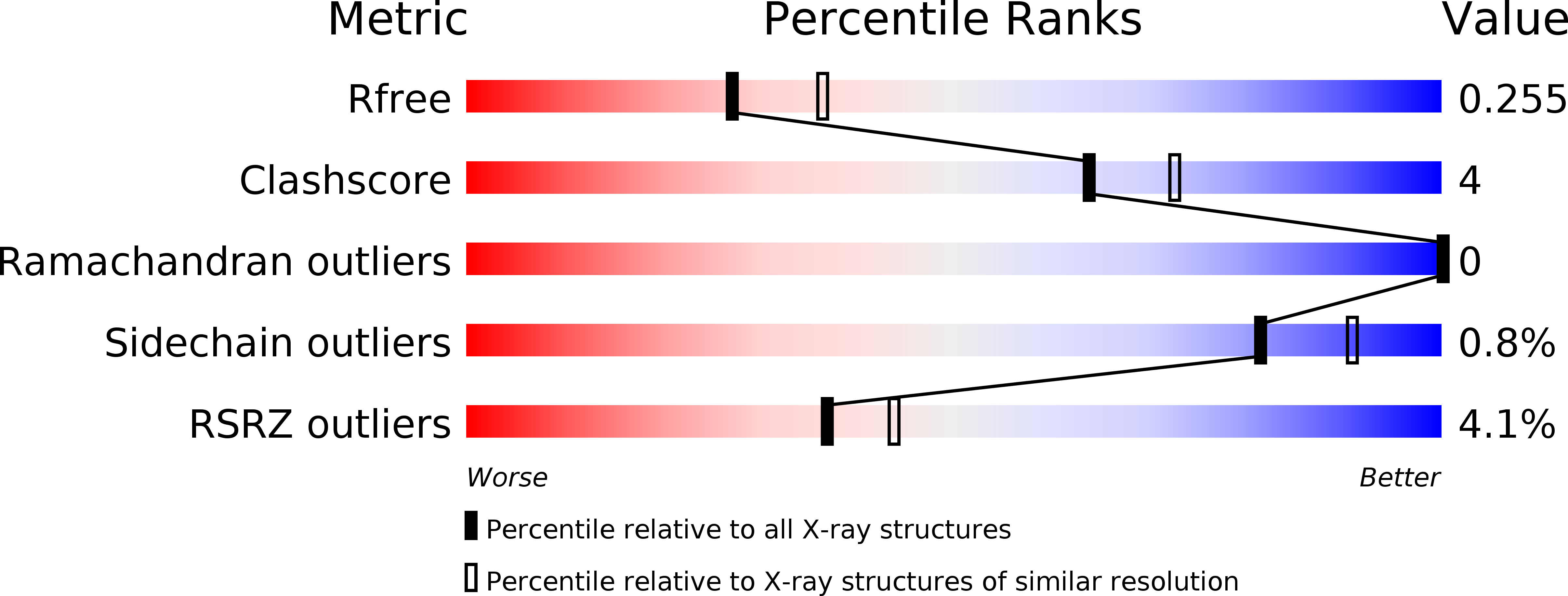
R-Value Free:
0.26
R-Value Work:
0.21
R-Value Observed:
0.21
Space Group:
C 1 2 1