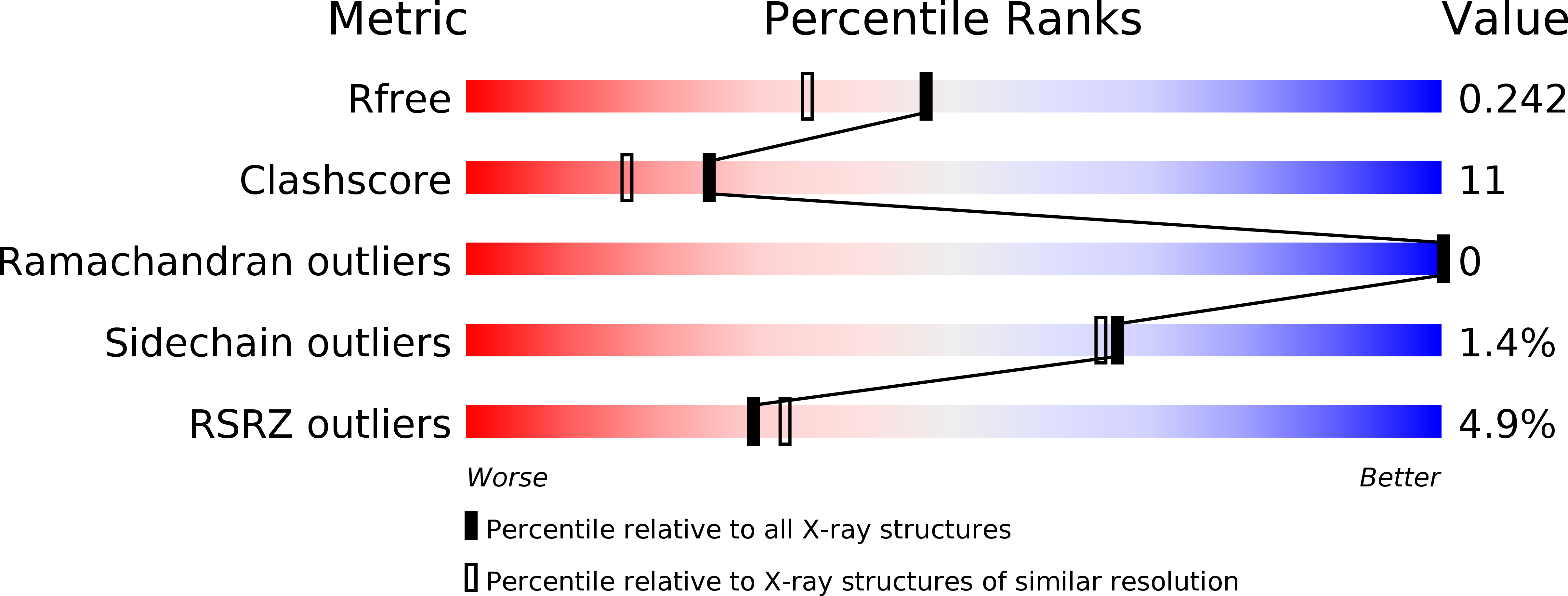
3N8B image
Deposition Date 2010-05-28
Release Date 2010-10-06
Last Version Date 2024-10-09
Entry Detail
PDB ID:
3N8B
Title:
Crystal Structure of Borrelia burgdorferi Pur-alpha
Biological Source:
Source Organism:
Host Organism:
Method Details:
Experimental Method:
Resolution:
1.90 Å
R-Value Free:
0.23
R-Value Work:
0.18
R-Value Observed:
0.18
Space Group:
I 21 21 21
Macromolecular Entities
Polymer Type:polypeptide(L)
Molecule:Uncharacterized protein
Gene (Uniprot):bpuR
Mutations:L17M, F27M, I64M
Chain IDs:A, B
Chain Length:98
Number of Molecules:2
Biological Source:Borrelia burgdorferi
Modified Residue
Compound ID Chain ID Parent Comp ID Details 2D Image
MSE A MET SELENOMETHIONINE
Primary Citation
Of bits and bugs--on the use of bioinformatics and a bacterial crystal structure to solve a eukaryotic repeat-protein structure.
Plos One 5 e13402 e13402 (2010)
PMID: 20976240 DOI: 10.1371/journal.pone.0013402

Abstact

Pur-α is a nucleic acid-binding protein involved in cell cycle control, transcription, and neuronal function. Initially no prediction of the three-dimensional structure of Pur-α was possible. However, recently we solved the X-ray structure of Pur-α from the fruitfly Drosophila melanogaster and showed that it contains a so-called PUR domain. Here we explain how we exploited bioinformatics tools in combination with X-ray structure determination of a bacterial homolog to obtain diffracting crystals and the high-resolution structure of Drosophila Pur-α. First, we used sensitive methods for remote-homology detection to find three repetitive regions in Pur-α. We realized that our lack of understanding how these repeats interact to form a globular domain was a major problem for crystallization and structure determination. With our information on the repeat motifs we then identified a distant bacterial homolog that contains only one repeat. We determined the bacterial crystal structure and found that two of the repeats interact to form a globular domain. Based on this bacterial structure, we calculated a computational model of the eukaryotic protein. The model allowed us to design a crystallizable fragment and to determine the structure of Drosophila Pur-α. Key for success was the fact that single repeats of the bacterial protein self-assembled into a globular domain, instructing us on the number and boundaries of repeats to be included for crystallization trials with the eukaryotic protein. This study demonstrates that the simpler structural domain arrangement of a distant prokaryotic protein can guide the design of eukaryotic crystallization constructs. Since many eukaryotic proteins contain multiple repeats or repeating domains, this approach might be instructive for structural studies of a range of proteins.

Legend

Protein

Chemical

Disease

Primary Citation of related structures