3N7K image
Deposition Date 2010-05-27
Release Date 2010-09-08
Last Version Date 2023-09-06
Entry Detail
PDB ID:
3N7K
Keywords:
Title:
Crystal structure of botulinum neurotoxin serotype C1 binding domain
Biological Source:
Source Organism:
Host Organism:
Method Details:
Experimental Method:
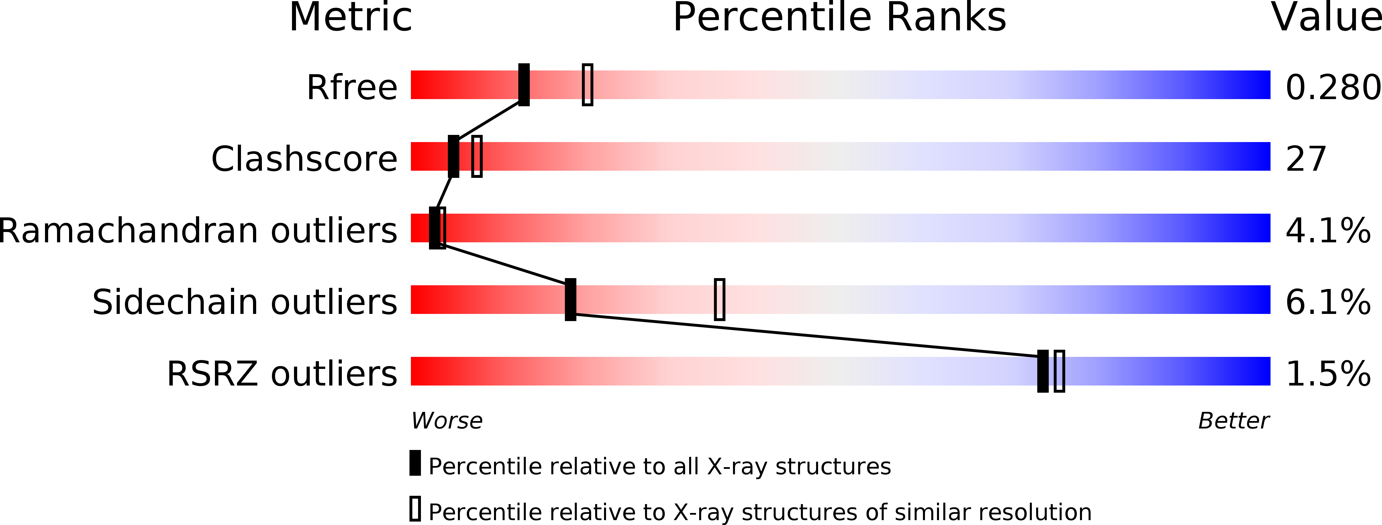
Resolution:
2.50 Å
R-Value Free:
0.28
R-Value Work:
0.22
R-Value Observed:
0.22
Space Group:
P 1 21 1
Macromolecular Entities
Polymer Type:polypeptide(L)
Molecule:Botulinum neurotoxin type C1
Chain IDs:A, B
Chain Length:426
Number of Molecules:2
Biological Source:Clostridium botulinum
Primary Citation
Identification of a Unique Ganglioside Binding Loop within Botulinum Neurotoxins C and D-SA .
Biochemistry 49 8117 8126 (2010)
PMID: 20731382 DOI: 10.1021/bi100865f

Abstact

The botulinum neurotoxins (BoNTs) are the most potent protein toxins for humans. There are seven serotypes of BoNTs (A-G) based on a lack of cross antiserum neutralization. BoNTs utilize gangliosides as components of the host receptors for binding and entry into neurons. Members of BoNT/C and BoNT/D serotypes include mosaic toxins that are organized in D/C and C/D toxins. One D/C mosaic toxin, BoNT/D-South Africa (BoNT/D-SA), was not fully neutralized by immunization with BoNT serotype C or D, which stimulated this study. Here the crystal structures of the receptor binding domains of BoNT/C, BoNT/D, and BoNT/D-SA are presented. Biochemical and cell binding studies show that BoNT/C and BoNT/D-SA possess unique mechanisms for ganglioside binding. These studies provide new information about how the BoNTs can enter host cells as well as a basis for understanding the immunological diversity of these neurotoxins.

Legend

Protein

Chemical

Disease

Primary Citation of related structures