3N5U image
Deposition Date 2010-05-25
Release Date 2010-08-11
Last Version Date 2024-02-21
Entry Detail
PDB ID:
3N5U
Title:
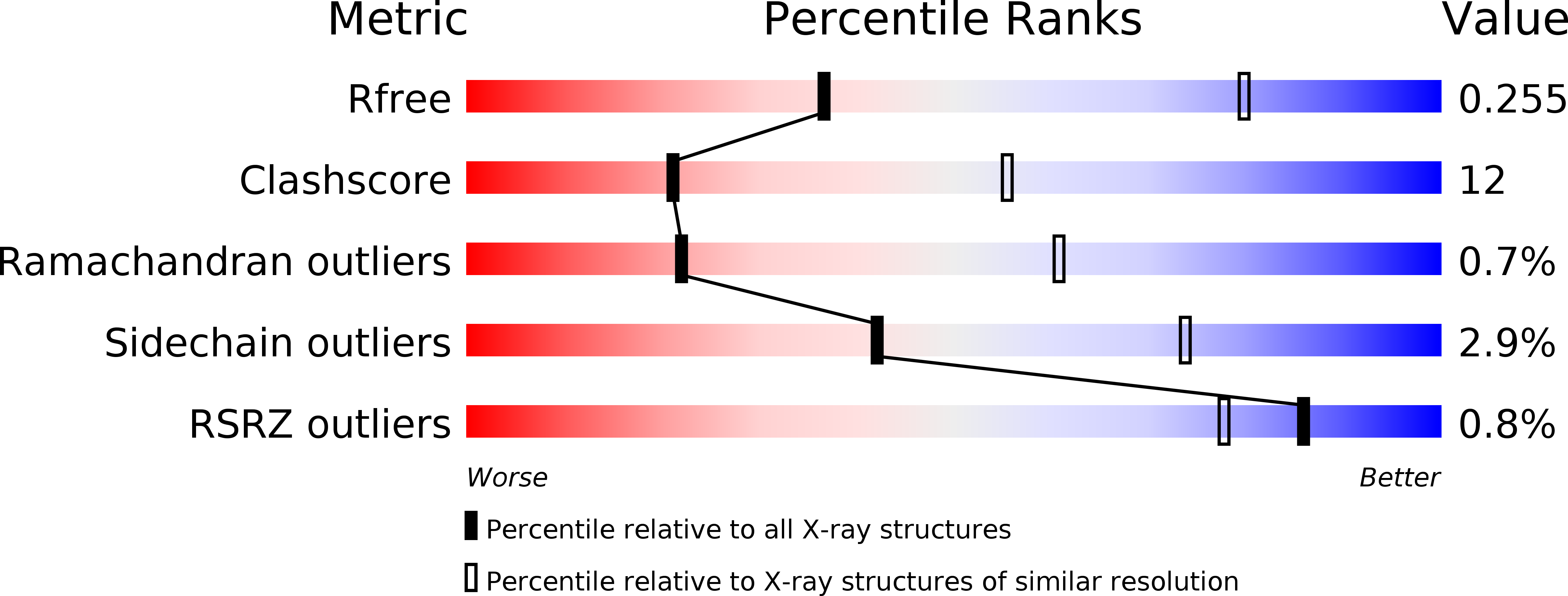
Crystal structure of an Rb C-terminal peptide bound to the catalytic subunit of PP1
Biological Source:
Source Organism:
Homo sapiens (Taxon ID: 9606)
Host Organism:
Method Details:
Experimental Method:
Resolution:
3.20 Å
R-Value Free:
0.26
R-Value Work:
0.22
R-Value Observed:
0.22
Space Group:
P 41 21 2
Macromolecular Entities
Polymer Type:polypeptide(L)
Molecule:Serine/threonine-protein phosphatase PP1-alpha catalytic subunit
Gene (Uniprot):PPP1CA
Chain IDs:A, B
Chain Length:300
Number of Molecules:2
Biological Source:Homo sapiens
Polymer Type:polypeptide(L)
Molecule:Retinoblastoma-associated protein
Gene (Uniprot):RB1
Chain IDs:C
Chain Length:13
Number of Molecules:1
Biological Source:Homo sapiens
Primary Citation
An overlapping kinase and phosphatase docking site regulates activity of the retinoblastoma protein.
Nat.Struct.Mol.Biol. 17 1051 1057 (2010)
PMID: 20694007 DOI: 10.1038/nsmb.1868

Abstact

The phosphorylation state and corresponding activity of the retinoblastoma tumor suppressor protein (Rb) are modulated by a balance of kinase and phosphatase activities. Here we characterize the association of Rb with the catalytic subunit of protein phosphatase 1 (PP1c). A crystal structure identifies an enzyme docking site in the Rb C-terminal domain that is required for efficient PP1c activity toward Rb. The phosphatase docking site overlaps with the known docking site for cyclin-dependent kinase (Cdk), and PP1 competition with Cdk-cyclins for Rb binding is sufficient to retain Rb activity and block cell-cycle advancement. These results provide the first detailed molecular insights into Rb activation and establish a novel mechanism for Rb regulation in which kinase and phosphatase compete for substrate docking.

Legend

Protein

Chemical

Disease

Primary Citation of related structures